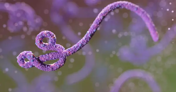
Ebola virus disease (EVD) is a highly infectious and often fatal illness caused by the Ebola virus. Currently, there is no specific treatment for EVD and the only known way to control outbreaks is through early detection and isolation of infected individuals, contact tracing, and infection prevention and control measures.
Researchers have discovered a cellular pathway that prevents the Ebola virus from exiting human cells, which could lead to the development of new antivirals. Viruses have developed strategies to initiate and sustain infection in their evolutionary battle for survival. The Ebola virus, for example, once inside a host cell, hijacks molecular pathways to replicate itself and eventually make its way back out of the cell into the bloodstream, where it can spread further.
But, in the case of Ebola and many other viruses, our own cells have defenses. A team led by scientists from the University of Pennsylvania School of Veterinary Medicine discovered a way to prevent the Ebola virus from exiting human cells in a study published in the Proceedings of the National Academy of Sciences.
An interaction between viral and host proteins prompts host cells to ramp up activity of a pathway responsible for digesting and recycling proteins, the team found. This activity, known as autophagy “self-eating,” allows fewer viral particles to reach the surface of a host cell, thus reducing the number that can exit into the bloodstream and further propagate infection.
This interaction appears to be part of an innate defense mechanism. A key Ebola virus protein appears to be specifically targeted by human cells and directed into the autophagy pathway, which is how cells process and recycle waste.
Ronald N. Harty
“This interaction appears to be part of an innate defense mechanism,” says Ronald N. Harty, a Penn Vet professor and the study’s senior author. “A key Ebola virus protein appears to be specifically targeted by human cells and directed into the autophagy pathway, which is how cells process and recycle waste.”
Harty’s lab has long been interested in the interaction between the viral protein VP40, which is found in both Ebola and Marburg viruses, and various human proteins. Previous research by the group discovered that a region of VP40 known as a PPXY motif binds to corresponding motifs known as WW domains on specific host proteins.
This PPXY-WW interaction frequently causes more viral particles to exit the cell, a process known as “budding.” However, when Harty and postdoc Jingjing Liang, the study’s lead author, screened various host proteins thought to play a role in the process, they discovered some that did the opposite when bound to VP40, causing budding to decrease. One of these was a protein called Bag3, about which they published a paper in PLOS Pathogens in 2017.
Despite the fact that Ebola is a potentially lethal virus, Harty and colleagues can study its mechanisms in a Biosafety Level 2 laboratory using virus-like particles (VLPs) that express VP40 instead of the virus itself. These VP40 VLPs are not infectious, but they can bud from host cells just like the real thing.
The Penn Vet researchers and colleagues from the Texas Biomedical Research Institute dug deeper in the new study to learn more about the mechanism by which Bag3 reduced budding. Bag3 is a “co-chaperone” protein, meaning it forms a complex with other proteins and chaperones them on their way to be digested, eventually in organelles called autolysosomes, as part of the autophagy process. Harty’s group used VP40 VLPs to confirm that VP40 bound to Bag3 and formed the protein complex. When the researchers added a compound known to inhibit the formation of this complex, they observed the release of VP40, which increased VLP budding activity.
To follow the activity of VP40 in real time, the team used powerful confocal microscopy, labeling each actor of interest with a different fluorescent tag. They observed that Bag3 was involved in sequestering VP40 in vesicles in the cell that would go on to undergo autophagy. Stuck in these vesicles and destined for the cellular “recycling center,” VP40 was unable to move to the cell membrane and bud.
“I think one of the most interesting things that we showed is the selectivity of the cargo,” Liang says. “We show that autophagy doesn’t just happen passively. Bag3 acts through the PPXY-WW interaction to specifically target VP40 to undergo autophagy.”
When the researchers added the autophagy-boosting drug rapamycin, VP40 sequestration increased while VLP budding decreased. Rapamycin works by inhibiting the activity of a pathway governed by a protein complex called mTORC1, a cellular sensor that activates protein synthesis when a cell requires raw material to grow. The researchers discovered that this pathway appeared to be important in regulating Ebola infection; in experiments with live virus conducted in a Biosafety Level 4 laboratory, they discovered that the virus could activate mTORC1 signaling, causing the cellular “factory” to produce materials the virus would need to expand and spread. In contrast, inhibiting mTORC1 with rapamycin directed the virus toward the autophagy pathway, where it would be digested by the cell’s autolysosomes.
“The virus wants the cell to grow, so it activates mTORC1,” Harty explains. “Autophagy does the opposite, balancing the cellular materials.” Autophagy is essential for normal cellular processes because it prevents the cell from becoming clogged with unnecessary or misfolded proteins and other materials floating around. However, this research suggests that the body can use autophagy to defend itself against harmful infections.
“We believe this is part of an arms race between our bodies and the virus,” Liang says. “Because the virus wants to shape its environment for its own survival, it evolved to manipulate mTORC1. However, the cell can use this pathway to defend itself against viral infection.”
The researchers hope that by learning more about the human body’s innate defenses against Ebola, they will be able to determine whether autophagy plays a role in other hemorrhagic viral infections, such as those that cause Marburg and Lassa fever. While the current experiments were mostly done on human liver cell lines, the researchers would like to see if autophagy and the mTORC1 pathway are involved in viral defense in other cell types, such as immune system macrophages, which are the primary cells involved in infection propagation.
Finally, Harty, Liang, and colleagues hope to discover as many viral vulnerabilities as possible, which will help inform drugs that could be part of a therapeutic cocktail, each targeting a different stage of infection, from viral entry to viral exit.
“This all contributes to our overall goal of understanding viral-host interactions and, by doing so, working to intervene to slow or stop infection,” Harty says.