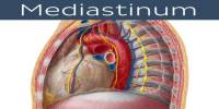
Sella Turcica
Definition
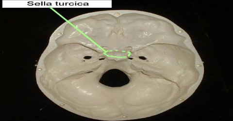
Sella Turcica is a bony cavity housing the pituitary gland, which is located in the brain and responsible for secreting a number of hormones. It serves as a cephalometric landmark. It includes the tuberculum sellae anteriorly and the dorsum sellae posteriorly; with its covering of dura mater it constitutes the hypophysial fossa that accommodates the hypophysis or pituitary gland.
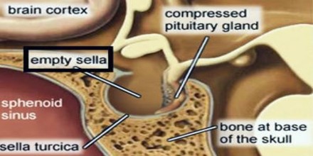
If the pituitary gland is revealed as absent from this cavity on imaging, scanning, or MRI examination, the condition is sometimes referred to as empty sella syndrome. When the pituitary gland is, in fact, flattened against the surrounding bone, a condition which nevertheless does not impede its function in the majority of cases.
Structure and Fnction of Sella Turcica
Sella turcica resembles as saddle-like depression, which provide place for the pituitary gland. Anatomically, the sella turcica has been expressed as variable. It is divided into three fragments and consists of an anterior wall, a floor, and a posterior wall.
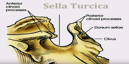
The posterior surface of attached with membrane, which is termed as dorsum sellae amd the anterior order is attached with the tuberculum sellae. The whole sellar area is made up of
- Optic Chiasma
- Pituitary Gland
- Internal Carotids
- Cavernous sinusand cranial nerves
- Sphenoid Sinuses
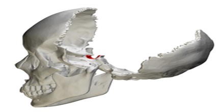
The cranial base of the skull is supported by sphenoid bone, which is looks like a butterfly. Sella turcica, cross-sectional depression present at the mid line of the sphenoid bone. The exact location can be described as sella turcica present just at the back of the void space identified as the sphenoid sinus, which sits it in the region of the center of the base of the cranium.
Completing the formation of the saddle posteriorly is the dorsum sellae, which is continuous with the clivus, inferoposteriorly. The dorsum sellae is terminated laterally by the posterior clinoid processes. Sella turcica providing the proper place to hold and support the pituitary gland.
Clinical Significance of Sella Turcica
Any anomaly or pathological condition in the pituitary gland can apparent from a distorted form of sella turcica, to a disorder in the malfunctioning of the secretion of the hormones secreted by the pituitary, which includes growth hormones (GH), thyroid stimulating hormone (TSH), prolactin, follicular stimulating hormone (FSH), and others.
Since the sella turcica forms a bony caudal border for the pituitary gland, a pituitary tumor usually extends upward in the rostral direction into the suprasellar region. This can result in compression of the optic chiasm, which lies on top of the pituitary, enveloping the pituitary stalk. Compression of the optic chiasm can lead to bitemporal hemianopsia, and, when there is no relevant trauma, this clinical finding is pathognomonic for a pituitary tumor.
Some pituitary adenomas can extend inferiorly, growing downward and invading the sphenoid bone and cavernous sinus. Adenomas greater than 10mm (macroadenomas) can cause remodeling of the underlying sphenoid bone altering the shape of the sella turcica.
Sella turcica is also usually used as a reference point with nasion to establish the base of the skull in cephalometric analysis. This is commonly done prior to orthodontic treatment.
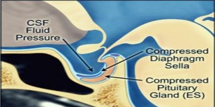
Empty sella defined as an enlargement or deformity of sella turcica that is completely or partially filled with CSF (cerebrospinal fluid). In fact, the expression ’empty sella’ is a not accurate, as the sella is not entirely vacant. The pituitary is constantly present both anatomically and physiologically, though it is dislocated downwards and squeezed by the pressure of cerebrospinal fluid. The different causes associated with Empty sella are as follows:
- Congenital nonappearance of the diaphragma sellae
- Incomplete formation of diaphragma sellae
- Expansion of the suprasellar subarachnoid space.
An empty sella is often a subsidiary anatomical shift and sporadically it consequences in anomalous pituitary function. Despite of the volume, the sella is partially or completely packed with cerebrospinal fluid. It can arise at any age and is not a gender specific condition, though the incidence rate is more in women with increasing age.
Sella turcica is also usually used as a reference point with nasion to establish the base of the skull in cephalometric analysis. This is commonly done prior to orthodontic treatment.