Contrast agents are drugs that boost the information content of diagnostic images. They improve the sensitivity and specificity of diagnostic images by changing the intrinsic properties of tissues, which influence the fundamental contrast mechanisms. The agent’s strategic localization can alter tissue properties and result in preferential enhancement. MRI is distinct from other diagnostic modalities in that it employs more than one intrinsic property of the tissue being imaged.
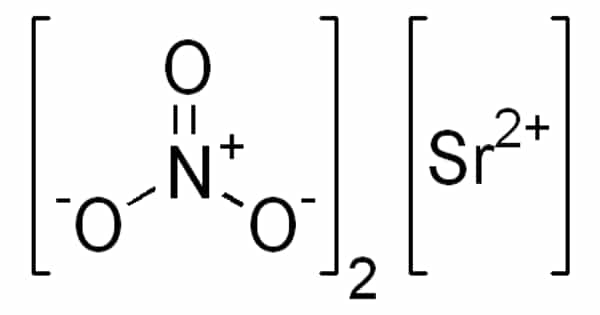
MRI contrast agents are contrast agents that are used in magnetic resonance imaging to improve the visibility of internal body structures (MRI). Gadolinium-based compounds are the most commonly used contrast enhancers. Following oral or intravenous administration, these MRI contrast agents reduce the relaxation times of nuclei within body tissues.
You are free to keep your best guesses. Engineers at Rice University’s George R. Brown School of Engineering are learning more about what happens when contrast agents are injected into your body for an MRI scan.
A Rice-led team delves deeper in a new study that could lead to better scans using molecular simulations that, unlike previous models, make no assumptions about the basic mechanisms at work when gadolinium agents are used to highlighting soft tissues.
Going back 40 years, in the NMR field people assumed liquid water is just a collection of marbles moving about, and the dipoles in the marbles randomly reorient. What Thiago does with his explicit simulation is show how the water network evolves in time. These are complicated, computationally intensive calculations.
Dilip Asthagiri
The study led by Rice chemical and biomolecular engineer Philip Singer, former associate research professor Dilip Asthagiri, now of Oak Ridge National Laboratory, and graduate student Thiago Pinheiro dos Santos appears in Physical Chemistry Chemical Physics.
It employs the sophisticated models first developed at Rice for oil and gas studies to conclusively analyze how hydrogen nuclei at body temperatures “relax” under nuclear magnetic resonance (NMR), the technology used by magnetic resonance imaging, aka MRI.
Doctors use MRI to “see” the state of soft tissues, including the brain, in a patient by inducing magnetic moments in the hydrogen nuclei of water molecules to align with the magnetic field, a process that can be manipulated when gadolinium agents are in the vicinity. The device detects bright spots when the aligned nuclei relax back to thermal equilibrium following an excitation. The faster they relax, the brighter the contrast.
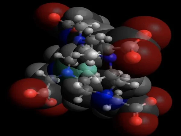
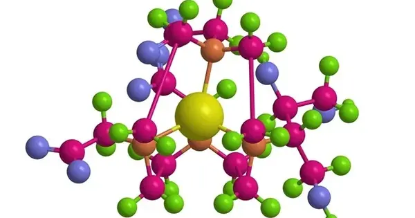
Gadolinium molecules are naturally paramagnetic and sensitive to magnetic excitation. Because they’re toxic, they are usually chelated when part of a contrast agent. “A chelate basically hugs the gadolinium and protects your body from directly interacting with the metal,” Pinheiro dos Santos said. “We’re asking, exactly how do these molecules behave?”
Though gadolinium-based contrast agents are injected by the ton into patients each year, how they work on a molecular level has never been fully understood.
“Going back 40 years, in the NMR field people assumed liquid water is just a collection of marbles moving about, and the dipoles in the marbles randomly reorient,” Asthagiri said. But such assumptions are limiting, he said. “What Thiago does with his explicit simulation is show how the water network evolves in time,” Asthagiri said. “These are complicated, computationally intensive calculations.”
The Rice simulations make use of highly refined, polarizable force fields to study the phenomenon in detail, and that required intensive GPU-accelerated computing. The team validated its molecular dynamics approach with experimental data by co-author Steven Greenbaum, a professor of physics at Hunter College in the City University of New York, whose lab specializes in NMR measurements of ionic and molecular transport processes in condensed matter.
The simulations revealed distinct differences in how the inner and outer shells of water molecules around gadolinium respond to thermal excitation. “The inner shell is the group of eight or nine water molecules around gadolinium,” Pinheiro dos Santos said. “They’re strongly attached to the gadolinium and they stay there for a long time, a few nanoseconds. The outer shell encompasses all of the remaining water molecules.”
The researchers found that while the structure of the inner shell does not change between 41 and 98.6 degrees Fahrenheit, its dynamics are very susceptible to thermal effects. They also discovered that temperature greatly affects the self-diffusivity of molecules in the gadolinium-water simulations in a way that affects outer-shell relaxation.
“Overall, these discoveries open a new way to elucidate how contrast agents respond at human body conditions during an MRI scan,” Singer said. “By better understanding this, one can develop new, safer, and more sensitive contrast agents, as well as use simulations to enhance the interpretation of MRI data.” He said future studies will examine chelated gadolinium complexes in fluids that are more representative of cellular interiors.