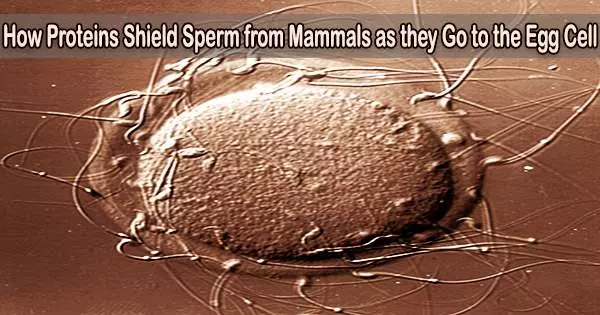
A range of proteins released by the accessory sex glands are present in mammalian seminal fluid and are crucial for the processes involved in fertilization. The spermadhesin AQN-3 is one of these proteins, which is present in ungulates and in particularly high concentrations in boars.
The protein was examined by a scientific team from the Leibniz Institute for Molecular Pharmacology, the Humboldt University of Berlin, and the Leibniz Institute for Zoo and Wildlife Research (Leibniz-IZW) and unexpected properties were found that may aid sperm in maintaining function up until they reach the egg. The scholarly journal Chemistry and Physics of Lipids publishes the findings.
The survival of male germ cells and their species-specific interactions with the elements of the female genital tract depend on the proteins in seminal fluid. In ungulates, there are five distinct spermadhesins present in the seminal fluid. Porcine semen contains exceptionally high concentrations of spermadhesins.
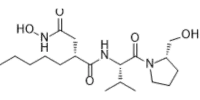
The spermadhesins AWN and AQN-3, named after the letter code of the first three amino acids of the protein sequence, have so far been synthesized in bacteria in order to better understand the function of these proteins, which, as their name suggests, attach themselves to the surface of sperm.
In a previous study, the scientific team led by Karin Mueller and Beate Braun from the Leibniz-IZW showed that AWN binds specifically to negatively charged lipids.
The pB1 protein belongs to a group of proteins that are known to bind to the phosphatidylcholine on the outside of the cell membrane in bovine sperm and thus have a stabilizing effect.
Peter Müller
These are mainly located on the cell membrane portion that faces the interior of the sperm cell, though. This raises the question of how the spermadhesins attach to the surface of the sperm?
Since it is known from work of other scientific groups that AWN forms aggregates with AQN-3, it was now investigated whether AQN-3 binds to typical lipids on the outside of the sperm membrane, such as phosphatidylcholine and sphingomyelin.
“Surprisingly, the applied model systems with lipid strips and vesicles showed that AQN-3 also binds selectively with negatively charged lipids, such as phosphatidic acid and various phosphatidylinositol phosphates,” explains Mueller.
Additionally, AQN-3 has a great propensity to combine with other proteins as well as with itself. Other research teams have identified the protein pB1 in the native AWN and AQN-3 aggregates in seminal fluid as yet another protein.
“The pB1 protein belongs to a group of proteins that are known to bind to the phosphatidylcholine on the outside of the cell membrane in bovine sperm and thus have a stabilizing effect,” says Peter Müller from the Humboldt University in Berlin.
Therefore, it is believed that pB1 forms aggregates to bind the protective protein envelope of AWN and AQN-3 to the sperm cell membrane. During fertilization, negative-charged lipids like phosphatidylinositol phosphates act as signaling molecules among other things. They may be “intercepted” by spermadhesins if they are generated and released too early, delaying the time the sperm reach the egg at the point of fertilization.
Interestingly, only pig species have spermadhesins as the primary constituent of their seminal fluid since the protein sheath around sperm is species-specific. This begs the questions of how their function is carried out in other species and whether assisted reproduction techniques could be used to take advantage of their protective qualities in the interest of species preservation.