With the aid of a brand-new laser technique known as FLUCS (Focused Light-induced Cytoplasmic Streaming), it is now able to specifically direct and affect motions within living cells and embryos.
Rapp OptoElectronic has now obtained a license for the technology created at the Max Planck Institute of Molecular Cell Biology and Genetics, which can aid in our understanding of illnesses affecting embryonic development.
Future cell biology and medical research will benefit from FLUCS as an add-on module for high-resolution microscopes, and it will also expand the field of microfluidics.
To see and examine biological processes in cells, sophisticated imaging techniques are utilized in cell biology and medical research. Understanding processes and causal linkages requires careful targeting of cells in controlled environments.
In order to examine the consequences of changing one component of a cell on internal mechanisms and relationships, researchers are consequently dependent on efficient tools.
However, a major issue with conventional methods of cell manipulation is that the modification disturbs the sample, which leads to misleading results. For the first time, the novel FLUCS approach now enables non-invasive cell manipulation, for use in developmental biology, for instance.
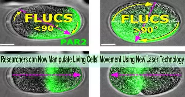
With the use of laser beams, the photomanipulation technique FLUCS enables precise influence and control over movements within cells and embryos. The beam selectively induces a thermal field in the cytoplasm. Due to the fast moving laser point, this locally alters the density and viscosity of the liquid medium and generates a flow.
We are pleased that the successful cooperation between the Max Planck Institute for Molecular Cell Biology and Genetics and Rapp OptoElectronic will bring first-class commercial products to the market that are far superior to the current state of the art. FLUCS makes microscopy interactive and opens up new possibilities for a variety of research areas.
Bernd Ctortecka
As opposed to traditional techniques like optical tweezers, the biomolecules floating in the cytoplasm are immediately set in motion without the need to change the sample. They can still freely interact with their surroundings. The technique can be utilized in particular to provide answers to significant queries regarding embryonic development.
A research team led by Moritz Kreysing from the Max Planck Institute (now at the Karlsruhe Institute of Technology) was able to generate controlled currents in living worm embryos and transport biomolecules to different parts of the growing embryo.
They were able to investigate the significance of cytoplasmic movement for the polarization of oocytes through controlled redistribution, raising the issue of which molecules must move precisely where throughout development.
A market-ready product
As part of a development cooperation, the FLUCS technology was transferred from the Max Planck Institute to the company Rapp OptoElectronic. Based on the successful joint development and the license agreement that has now been concluded, Rapp OptoElectronic is offering FLUCS as a market-ready product to researchers and industrial users worldwide.
A pilot system is located in the LMF (Light Microscopy Facility) of the Max Planck Institute of Molecular Cell Biology and Genetics in Dresden. Here, FLUCS is available to interested scientists from inside and outside the Max Planck Society for their research.
The tool is easily utilized for photo modification since it is easily integrated as an add-on module to high-resolution microscopes via standard interfaces.
“FLUCS fills a gap in the previously available micro-manipulation techniques to study the causes and consequences of intracellular movement. Directed liquid flows are induced by moderately warming up the sample with a laser spot. Their path can be easily specified individually using the user-friendly software, for example as a line, circle or free form. In this way, cell components such as organelles, PAR proteins and even chromatin can be moved freely in the cell nucleus without having to hold or fix them,” says Sven Warnck, Managing Director of Rapp OptoElectronic.
Diverse applications
The possible applications are diverse. In cell biology, for instance, it is possible to reverse PAR proteins and so affect embryonic development by using artificially produced cytoplasmic currents.
For instance, molecular mechanisms and signaling pathways in cells can be further studied in medical research to aid in the development of new medications. By using FLUCS in microfluidics, it is possible to analyze the behavior of liquid quantities in the micro or picoliter range in more detail, advancing new approaches to laboratory measurement technology, quality control, and food safety.
“We are pleased that the successful cooperation between the Max Planck Institute for Molecular Cell Biology and Genetics and Rapp OptoElectronic will bring first-class commercial products to the market that are far superior to the current state of the art. FLUCS makes microscopy interactive and opens up new possibilities for a variety of research areas,” says Bernd Ctortecka, patent and license manager at Max Planck Innovation, the technology transfer organization of the Max Planck Society.