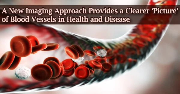
Researchers at Johns Hopkins Medicine have developed and tested a new imaging approach that they claim can speed up imaging-based research in the lab by allowing researchers to acquire images of blood arteries at various spatial scales.
The “VascuViz” technology, which has been tested in animal tissues, uses a quick-setting polymer combination to fill blood arteries and make them visible in several imaging modalities. According to the researchers, the method allows them to view the structure of a tissue’s vasculature, which, when combined with comprehensive mathematical models or complementary images of other tissue parts, can help to understand the complicated role of blood flow in health and sickness.
They claim that the combined images of blood vessels will help researchers better understand the biology of disorders involving aberrant blood flow, such as cancer and stroke, as well as the architecture and functions of tissues throughout the body.
The report published Feb. 10 in Nature Methods.
“Usually, if you want to gather data on blood vessels in a given tissue and combine it with all of its surrounding context like the structure and the types of cells growing there, you have to re-label the tissue several times, acquire multiple images and piece together the complementary information,” says Arvind Pathak, Ph.D., professor of radiology, biomedical and electrical engineering and member of the Sidney Kimmel Comprehensive Cancer at the Johns Hopkins University School of Medicine.
“This can be an expensive and time-consuming process that risks destroying the tissue’s architecture, precluding our ability to use the combined information in novel ways.”
In the lab, researchers use a variety of imaging techniques to investigate the role of blood arteries, including MRI, CT, and microscopy. These images are helpful in understanding how tissues develop disease and respond to treatment.
Usually, if you want to gather data on blood vessels in a given tissue and combine it with all of its surrounding context like the structure and the types of cells growing there, you have to re-label the tissue several times, acquire multiple images and piece together the complementary information. This can be an expensive and time-consuming process that risks destroying the tissue’s architecture, precluding our ability to use the combined information in novel ways.
Arvind Pathak
However, combining the information included in various images has proven difficult due to the fact that agents employed to make a blood vessel visible to one imaging modality may render it invisible to another. The amount of data researchers may get from a single sample is thus limited.
VascuViz solves this difficulty by allowing researchers to acquire a comprehensive understanding of blood vessels and related tissue components with less time and effort by making the structure of the largest arteries to the smallest microvasculature visible using a variety of imaging technologies.
VascuViz’s creation is a hallmark of the expanding discipline of “image-based” vascular systems biology, and is particularly valuable in developing computerized visualizations of how complicated biological systems like the circulatory system work.
“Now, rather than using an approximation, we can more precisely estimate features like blood flow in actual blood vessels and combine it with complementary information, such as cell density,” says Akanksha Bhargava, Ph.D., postdoctoral fellow in the Pathak Lab within the Department of Radiology and Radiological Science at the Johns Hopkins University School of Medicine.
To do so, VascuViz-based measurements are fed into computer simulations of blood flow, such as those used by Bhargava in his cancer research. Bhargava examined multiple combinations of current imaging agents and their appropriateness for various imaging modalities in order to develop VascuViz.
She discovered that combining a CT contrast agent called BriteVu and a fluorescently labeled MRI contrast agent called Galbumin-Rhodamine to create a compound that makes the macro and microvasculature visible simultaneously when imaging with MRI, CT, and optical imaging techniques without interference after multiple iterations.
The researchers subsequently examined the molecule in a range of mice tissues, perfusing it through the circulatory system of breast cancer models, leg muscles, the brain, and kidney tissues while it was still operating in test tubes.
The MRI, CT, and optical microscope images of the tissues were then merged to create amazing 3D visualizations of the vasculature and accompanying components that make up these disease models and organ systems.
Pathak and his team anticipate that VascuViz will be widely embraced by scientists due to its cost and readily available components, allowing them to throw fresh light on a variety of disorders involving the vasculature.
Benjamin Monteagudo, Priyanka Kushwaha, Janaka Senarathna, Yunke Ren, Ryan Riddle, and Manisha Aggarwal of the Johns Hopkins University School of Medicine were also participated in this work.
This work was supported by the National Cancer Institute (51R01CA196701-05, 1R01CA237597-01A1), the National Institute of Dental & Craniofacial Research (5R01DE027957-02) and NIH Instrumentation grant (S10OD012287) and the Sidney Kimmel Comprehensive Cancer Center, Quantitative Sciences Pilot Project Grant.