X-rays are a form of electromagnetic radiation, with a wavelength shorter than that of visible light, which allows them to penetrate solid objects and produce images of bones and other internal structures.
Without harming the material, X-ray fluorescence examination enables elemental analysis in a range of settings. The elements distribution may be determined with greater accuracy the smaller the X-ray microbeam diameter.
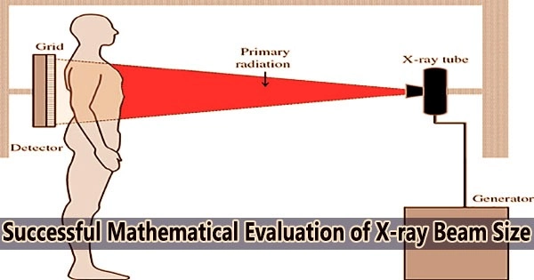
Because X-ray beams are not visible, an accurate method is needed to determine beam diameter. An X-ray beam actually refers to a stream of X-ray photons that are generated by an X-ray machine or other X-ray sources, such as radioactive isotopes.
A research group led by Professor Kouichi Tsuji and Specially Appointed Assistant Professor Tsugufumi Matsuyama of the Graduate School of Engineering, and Professor Hideyuki Ishi of the Graduate School of Science, at Osaka Metropolitan University, has developed a new method to evaluate the diameter of X-ray microbeams based on mathematical analysis.
We hope that this evaluation method will be widely used, and that the establishment of this method of evaluating spatial resolution in X-ray fluorescence will contribute to the development of a wide range of fields, including material development and bio-imaging.
Professor Kouichi Tsuji
It was discovered during the process of verifying the novel method that it can be used to compute beam sizes more precisely than the standard methods that were previously in use.
There isn’t yet a standard evaluation method for X-ray beam diameter. This evaluation technique is anticipated to be widely embraced as an international standard because it was developed utilizing mathematical analysis.
Materials development, environmental analysis, forensic science, biological sample analysis, and examination of archaeological and culturally significant artefacts are a few possible uses for X-ray fluorescence analysis.
Professor Tsuji concluded, “We hope that this evaluation method will be widely used, and that the establishment of this method of evaluating spatial resolution in X-ray fluorescence will contribute to the development of a wide range of fields, including material development and bio-imaging.”
The research results were published in X-Ray Spectrometry on January 9, 2023.