In biophotonic and biomedical applications, sensitive detection and imaging in the bio-microenvironment is highly desired. However, conventional photonic materials are inherently incompatible with and invasive to bio-systems. To address this issue, Chinese scientists reviewed recent advances in biophotonic probes such as bio-lasers, biophotonic waveguides, and bio-microlenses made from biological entities with inherent biocompatibility and minimal invasiveness, with applications in bio-detection and imaging. These biophotonic probes open up entirely new avenues of investigation for biophotonic research and biomedical applications.
The rapid advancement of biophotonics and biomedical sciences places a high demand on photonic structures capable of manipulating light at small scales for sensitive detection of biological signals and precise imaging of cellular structures in the bio-microenvironment. Unfortunately, when interacting with biological systems, conventional photonic structures based on artificial materials invariably exhibit incompatibility and invasiveness.
The design of biophotonic probes from abundant natural materials, particularly biological entities such as viruses, cells, and tissues, with the capability of multifunctional light manipulation at target sites can greatly increase biocompatibility while minimizing invasiveness to the biological microenvironment.
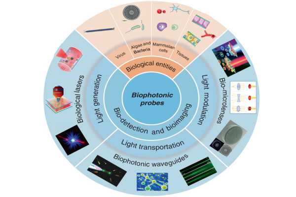
A team of scientists led by Professor Baojun Li and Professor Hongbao Xin from the Institute of Nanophotonics at Jinan University in China reviewed the intriguing progress of emerging biophotonic probes made from biological entities such as viruses, bacteria, cells, and tissues for bio-detection and imaging in a new paper published in Light Science & Application. They conducted a systematic review of three biophotonic probes with distinct optical functions: biological lasers for light generation, cell-based biophotonic waveguides for light transportation, and bio-microlenses for light modulation.
Sensitive detection and imaging in the bio-microenvironment are highly desired in biophotonic and biomedical applications. These biophotonic probes open up entirely new windows for biophotonic researches and biomedical applications.
Photonic probes’ potential biomedical applications require effective control and modulation of light generation in a variety of biochemical environments. In this regard, lasers’ unique properties, such as high intensity, directionality, and monochromatic emission, have made them one of the most useful tools in biomedical applications. Bio-lasers, as opposed to traditional laser devices, use biological entities such as cells, tissues, and viruses as part of the cavity and/or gain medium in a biological system. Cell lasers, tissue lasers, and virus lasers are the three types of bio-lasers. These bio-lasers avoid the biohazards of conventional laser devices.
Bio-lasers can serve as highly sensitive tools in a variety of biomedical applications, including cellular tagging and tracking, diagnostics, intracellular sensing, and novel imaging because their optical output is tightly related to the biological structures and activities of biological systems. Whispering gallery modes (WGM) microdisks with slightly different diameters, for example, produced clearly different lasing output spectra. Intracellular cell lasers created by incorporating these microdisks into cells allowed for the simultaneous tagging and tracking of individual cells from large cell populations.
Optical waveguides, in addition to bio-lasers for bio-detection and imaging in biological systems, play important roles in bio-microenvironments. Optical waveguides, as the primary component for light transportation, can deliver light signals in bio-microenvironments for further real-time analysis, and optical waveguides play an indispensable role in breaking the tissue penetration limit of light by transporting light into deep tissues. Living cells have enormous potential for in situ formations of biophotonic waveguides that are inherently elastic, biocompatible, and biodegradable, which could solve the problem of invasiveness and low biocompatibility of conventional materials-based optical waveguides.
Because the refractive index of biological cells (around 1.38) is slightly higher than that of water (around 1.33), light can be guided through a chain of cells via total internal reflection at the cell membrane-water interface. Optical trapping is a feasible and noninvasive method for assembling cell-based biophotonic waveguides. Biophotonic waveguides can be formed by assembling a chain of bacteria cells using laser light launched by a tapered optical fiber.
Light can travel through cell chains as long as tens of microns. In another case, nonlinear optical effects have been used to form biophotonic waveguides based on living cells such as algae and red blood cells (RBCs), resulting in stable long-distance light propagation with low loss in biological environments. These cell-based biophotonic waveguides can be used as a biophotonic probe for cell imaging and detection of biological microenvironments. Biophotonic waveguides formed by RBCs, for example, offer a potential detection technique for blood pH sensing and diagnosis of blood-related disorders.
Another important optical device for light modulation is the optical lens. Surprisingly, some living biological cells, acting as bio-microlenses, can confine light in biological systems. Cyanobacteria, for example, act as spherical microlenses, confining light into a focal spot near the plasma membrane on the backside of the light source. Many mammalian cells exhibit lensing behavior at a higher level of cellular complexity. RBCs are a type of disk-shaped microstructured envelope that can be used as adaptable bio-microlens due to their inherent deformability and lack of nucleus and organelles.
Because morphological abnormalities of RBCs are closely related to blood-related diseases, RBCs with biosensing properties can be used as a noninvasive, label-free, and rapid screening tool to distinguish abnormal RBCs from healthy cases. Biological cells have also been used as biomagnified to image living cells or other nanostructures without using labels.
These biophotonic probes open up completely new avenues for biophotonic research as well as biomedical applications, such as bio-lasers for bio-detection, cell tagging, and tissue imaging, biophotonic waveguides based on living cells for optical detection and sensing, and bio-microlenses for single-cell imaging and blood diagnostics. These biophotonic probes have numerous advantages over conventional photonic components.
In comparison to traditional synthetic materials, they have inherent and favorable opportunities for biocompatibility and biodegradability. Furthermore, the development of biophotonic probes based on biological cells/tissues allows these biological entities to serve as optical components as well as testing samples, allowing for in vivo and real-time sensing, detection, and imaging.
Despite significant progress, the authors emphasize that the overall development of biophotonic probes is still in its infancy, with much more to be discovered. They noted that more research is needed to fully comprehend and discover the vast and diverse family of living organisms that can serve as photonic probes. Furthermore, as proof of concept, most concepts and techniques have been demonstrated by in vitro or animal studies thus far.
Much more research is needed in the future to demonstrate the feasibility of preclinical and clinical practice applications. They also suggested that biophotonic probes, such as bio-microlenses, integrated into a smartphone-based platform have great potential in bio-detection, imaging, and molecular diagnosis with clinical samples in a portable and real-time manner, which is critical in resource-limited areas.
