Scientists can now create high-resolution photographs of the human spinal cord during surgery, thanks to technology developed at UC Riverside. The development could provide real relief to millions of people suffering from persistent back pain.
The technology, known as fUSI or functional ultrasound imaging, allows clinicians to not only visualize the spinal cord but also track its reaction to different treatments in real time. A study published in the journal Neuron describes how fUSI worked for six persons who received electrical stimulation for chronic back pain management.
“The fUSI scanner is freely mobile across various settings and eliminates the requirement for the extensive infrastructure associated with classical neuroimaging techniques, such as functional magnetic resonance imaging (fMRI),” said Vasileios Christopoulos, assistant professor of bioengineering at UCR who helped develop the technology. “Additionally, it offers ten times the sensitivity for detecting neuroactivation compared to fMRI.”
The fUSI scanner is freely mobile across various settings and eliminates the requirement for the extensive infrastructure associated with classical neuroimaging techniques, such as functional magnetic resonance imaging (fMRI).
Vasileios Christopoulos
Until now, it has been difficult to determine whether a back pain treatment is effective because patients are under anesthesia and unconscious. Thus, patients are unable to provide verbal input on their pain levels during therapy. “Using ultrasound, scientists can monitor changes in blood flow in the spinal cord caused by electrical stimulation. This could indicate that the treatment is effective,” Christopoulos said.
The spinal cord is a “unfriendly area” for standard imaging techniques because of significant motion artifacts, such as heart pulse and respiration. “These movements introduce unwanted noise into the signal, making the spinal cord an unfavorable target for traditional neuroimaging techniques,” Christopoulos told reporters.
By contrast, fUSI is less sensitive to motion artifacts. It emits sound waves into the area of interest, and red blood cells in that area echo the sound, producing a clear image. “It’s like submarine sonar, which uses sound to navigate and detect objects underwater,” Christopoulos said. “Based on the strength and speed of the echo, they can learn a lot about the objects nearby.”

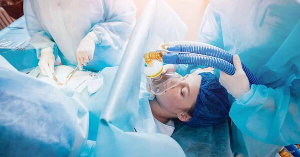
Christopoulos collaborated with the USC Neurorestoration Center at Keck Hospital to test the method on six chronic low back pain patients. These patients were previously scheduled for last-ditch pain surgery because no other therapies, including medications, had alleviated their misery. For this surgery, physicians stimulated the spinal cord using electrodes in the expectation that the voltage would relieve the patient’s pain and improve their quality of life.
“If you bump your hand, instinctively, you rub it. Rubbing increases blood flow, stimulates sensory nerves, and sends a signal to your brain that masks the pain,” Christopoulos said. “We believe spinal cord stimulation may work the same way, but we needed a way to view the activation of the spinal cord induced by the stimulation.”
The Neuron paper details how fUSI can detect blood flow changes at unprecedented levels of less than 1 millimeter per second. For comparison, fMRI is only able to detect changes of 2 centimeters per second.
“We have big arteries and smaller branches, the capillaries. They are extremely thin, penetrating your brain and spinal cord, and bringing oxygen places so they can survive,” Christopoulos said. “With fUSI, we can measure these tiny but critical changes in blood flow.”
Typically, this type of surgery has a 50% success rate. Christopoulos thinks that increased monitoring of blood flow variations will result in a significant increase in this rate. “We needed to know how fast the blood was flowing, how strong it was, and how long it took for blood flow to return to normal following spinal stimulation. “Now we’ll have these answers,” Christopoulos stated.
Moving forward, the researchers hope to demonstrate that fUSI can help optimize therapy for individuals who have lost bladder control due to spinal cord damage or aging. “We may be able to modulate the spinal cord neurons to improve bladder control,” Christopoulos told the audience.