In the central nervous system, microglia serve as the first line of defense, continually searching for pathogens and abnormalities and releasing tiny proteins called cytokines to fight infections.
Previous studies have demonstrated that microglia play a crucial role in transferring traumatic memories from short to long-term memory (fear memory consolidation) in mice that have been trained to dread a specific environment (contextual fear conditioning) and in the subsequent extinction of such memories.
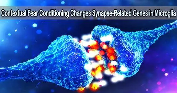
Researchers from Tohoku University have now shown that the consolidation and extinction of contextual fear conditioning alter the microglial genes linked to the synapse structures that enable neurons to communicate with one another.
This reveals the pathways connecting microglia and neuronal activity relevant to fear conditioning and suggests that microglia and neurons crosstalk via ‘non-immune’ roles.
Details of their research were published in the journal Brain Research Bulletin on August 18, 2022.
Dr. Zhiqian Yu and Professor Hiroaki Tomita from Tohoku University’s Graduate School of Medicine and their team has previously revealed that when mice were subjected to chronic and acute stress, their microglial released a type of cytokine known as TNF-a, which is used by the immune system for cell signaling.
Under contextual fear conditions, immune-related genes decreased with no significant morphological changes, indicating a change in microglial activation states. All of this suggests that microglia-neuron communication mediates contextual fear conditioning, and it may be based on non-immune functions.
Dr. Zhiqian Yu
TNF-a increased during the development of fear memories, whereas it decreased upon extinction. Additionally, TNF-a in the hippocampi prevents the consolidation and recall of spatial and contextual terror memories.
The scientists used microarray techniques in the microglia of mice that had undergone contextual fear conditioning, building on the findings of the prior study. They demonstrated that contextual fear conditioning alters the synapse-related genes in microglia.
However, they also found that the immunological dysfunction caused by consolidating the fear memory in microglia persisted even during the process of extinction.
Within the microglia’s plethora of synaptic function-related genes, Gamma-aminobutyric acid (GABA) and GABAR receptors (GABAR) are the earliest neurotransmitter systems to emerge during development. The GABARB3 encodes the ?3 subunit of GABAA receptors in neurological disorders such as epilepsy and autism.
“Using real-time PCR and immune stain technologies, we found that GABRB3 was expressed in microglial cytoplasm and the long branching processes of the hippocampus,” said Yu. “The mRNA and protein levels of GABRB3 changed significantly after fear memory consolidation but recovered after extinction.”
The researchers also looked at the Synapsin protein family, which controls neurotransmitter release at synapses. In MG-6 cell line and primary microglia that expanded during the consolidation of fear memories but recovered following the extinction of those memories, microglial Synapsin transcription was expressed.
“Under contextual fear conditions, immune-related genes decreased with no significant morphological changes, indicating a change in microglial activation states,” added Yu. “All of this suggests that microglia-neuron communication mediates contextual fear conditioning, and it may be based on non-immune functions,” said Yu.