A large-scale brain network refers to a complex web of interconnected brain regions that work together to perform various cognitive, sensory, and motor functions. The brain is a highly integrated system, with different regions communicating and coordinating their activities to produce behavior and cognition.
When we daydream or revisit memories, a large portion of our brain “lights up,” or becomes more active. The Default Mode Network (DMN) is so named because it is more active when the brain is not focused on the outside world.
The DMN has been related to a variety of brain illnesses, including Alzheimer’s, attention deficit/hyperactivity disorder, and mood disorders. However, the DMN’s neurophysiological basis is not well understood.
Neuroimaging techniques, such as functional magnetic resonance imaging (fMRI), cannot measure neural activity directly. To address this knowledge gap, a research team led by Ian Shih, PhD, professor and vice chair of the Department of Neurology and associate director of the Biomedical Research Imaging Center, has created a novel experimental platform that is able to optically record local neuronal activity during brain-wide fMRI in rodents.
“We hope that this work will pave the way for future translational studies aimed at controlling large-scale brain networks,” said Shih. “This could help design network-based treatment regimens for many neurological and neuropsychiatric disorders.”
This is important neuronal evidence highlighting the role of the anterior insular cortex in controlling DMN activity.
Professor Ian Shih
The study, which was published in the journal Science Advances, examined the dynamic activity of DMN-related brain regions and analyzed them with a variety of computational approaches.
The DMN is one of our brain’s large-scale brain networks. When we first learn about the brain, we are told that each component of the brain has a certain purpose. However, many brain areas activate and deactivate together during behavior and cognition, forming large-scale brain networks that function similarly to a team.
Understanding large-scale brain networks is critical for understanding the complex interplay between different brain regions and how they contribute to behavior and cognition.
As neuroscientists realize that some cognitive activities rely on “functionally connected” brain regions, they are becoming more interested in these large-scale networks. The DMN is active when a person is awake and at rest, such as when they are daydreaming, recovering memories, or imagining the future.
However, obtaining the neuronal data required to understand dynamic DMN activity in human subjects is difficult, so Shih and colleagues turned to an animal model to study the network, in which putative DMN-related brain regions have been identified.
“We used a rodent model, where genetically encoded calcium sensors were expressed in neurons,” said first author Tzu-Hao Harry Chao, PhD, who built and validated this experimental platform in the Shih lab. “This allowed us to record neuronal activity in multiple DMN-related brain regions by detecting changes in fluorescence via optical fibers without interfering with the measurement of fMRI signals.”
Fiber photometry is a technique that employs fiber optics to provide certain wavelengths of light in order to activate fluorescent proteins that respond to that wavelength and record activity-dependent light emission. Scientists can directly monitor activity from a specific population of cells or neurochemicals within a specific area of the brain using this method.
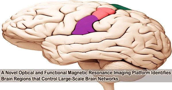
Using this novel experimental platform, Chao and colleagues demonstrated that activation of one area of the brain the anterior insular cortex is associated with suppressing, or “turning off,” the Default Mode Network.
The insular cortex is located in the cortex of the human brain and is “insulated” by the frontal, parietal, and temporal lobes. The insula is in charge of various critical brain activities, such as processing our five senses, managing hand-eye coordination, and self-awareness. The insula also plays a critical role in social and addiction-related behaviors.
“This is important neuronal evidence highlighting the role of the anterior insular cortex in controlling DMN activity,” Shih explained.
In collaboration with Vinod Menon, PhD, another senior author and professor of the Department of Psychiatry & Behavioral Sciences at Stanford University, the research team further used advanced computational approaches to identify the brain states and information flow during these conditions.
The team also discovered that the rodent cortex’s prelimbic area alternates its synchronization with the DMN and anterior insular cortex, implying that the rodent brain’s prelimbic cortex may also play a role in the salience network, another large-scale brain network important for attention, sensory processing, and goal-directed behavior.