Excessive folding of the gyri, or convolutions, in the human cerebral cortex can be caused by a condition called gyrification. Gyrification is a process that occurs during brain development, where the cerebral cortex becomes folded and creased to increase its surface area. This allows for more neurons to be packed into the same amount of space, which in turn can increase the brain’s processing power. Some genetic conditions that affect brain development, such as lissencephaly, can also cause excessive gyrification of the cerebral cortex. However, it is important to note that some excessive folding of the gyri can also be seen in certain neurological disorder such as Schizophrenia.
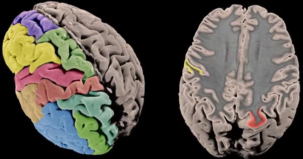
Researchers have discovered a mutation that causes excessive folding in the wrinkly cerebral cortex of the human brain, resulting in impaired cognitive function. The cerebral cortex, or outer layer of the human brain, controls cognitive and executive function, from conscious thought to speech to emotional control. It is distinguished by its distinctive gyri and sulci (ridges and furrows).
The cerebral cortex is made up of more than 10 billion cells and 100 trillion connections in a layer of gray matter that is only five millimeters thick – about the thickness of three stacked quarters.
Most animals with large brains have cortical folding, which allows a large area of cerebral cortex tissue (about 2.6 square feet) to be compacted inside the skull. The more cortical folding there is, the more advanced and complex the species’ cognitive functions are. Lower-order species, such as mice and rats, have smaller, smoother-surfaced brains, whereas higher-order species, such as elephants, porpoises, and apes, have varying degrees of gyrification or folding of the cerebral cortex. Humans have among the most wrinkly brains, which is thought to be an indicator of advanced evolution.
Once we determined that TMEM161B was the culprit, we set out to figure out how excessive folding happens. We discovered that the protein regulates the cellular skeleton and polarity, which in turn regulate folding.
Lu Wang
In some humans, however, excess folding of the cerebral cortex is associated not with greater cognitive abilities, but the opposite and linked to neurodevelopmental delay, intellectual disability and epileptic seizures. The genes controlling this folding are mostly unknown.
Writing in the issue of PNAS, researchers at the University of California San Diego School of Medicine and Rady Children’s Institute for Genomic Medicine describe new findings that deepen understanding of human gyrification.
An international consortium of researchers called the Neurogenetics Consortium, led by senior study author Joseph Gleeson, MD, Rady Professor of Neuroscience at UC San Diego School of Medicine and director of neuroscience research at the Rady Children’s Institute for Genomic Medicine, performed genomic analysis on nearly 10,000 families with pediatric brain disease over the course of ten years to look for new causes of disease.
“From our cohort, we found four families with a condition called polymicrogyria, meaning too many gyri that are too tightly packed,” said Gleeson. “Until recently, most hospitals treating patients with this condition did not test for genetic causes. The Consortium was able to analyze all four families together, which aided in our discovery of a cause for this condition.”
All four families had mutations in the Transmembrane Protein 161B (TMEM161B) gene, which produces a protein with an unknown function on cell surfaces.
“Once we determined that TMEM161B was the culprit, we set out to figure out how excessive folding happens,” said first author Lu Wang, Ph.D., a postdoctoral fellow in the Gleeson lab. “We discovered that the protein regulates the cellular skeleton and polarity, which in turn regulate folding.”
The researchers discovered defects in neural cell interactions early in embryogenesis using stem cells derived from patient skin samples and engineered mice.
“We discovered that the gene is both necessary and sufficient for the cytoskeletal changes required for neural cells to interact with one another,” Wang explained. “The fact that the gene first appeared in evolution in sponges, which don’t even have a brain, suggests that the protein must have other functions. We discovered that this protein plays an important role in regulating the number of folds in the human brain.”
The study’s authors emphasized the importance of genetic discovery studies in determining the causes of human disease, but that these discoveries can take many years to evolve into new treatments. “We hope that physicians and scientists will build on our findings to improve the diagnosis and care of patients with brain disease,” Gleeson said.