SARS-CoV-2, the virus that causes COVID-19, is more stable and slower changing than the earlier variant that caused the SARS outbreak in 2003, according to new computational simulations of the behavior of SARS-CoV-1 and SARS-CoV-2 spike proteins prior to fusion with human cell receptors.
SARS-CoV-1 and SARS-CoV-2 (severe acute respiratory syndrome coronaviruses 1 and 2) show striking similarities, and researchers aren’t sure why the latter has proved more contagious.
They have identified the spike proteins of each as a potential source of the varied transmissibility via binding to the host cell angiotensin-converting enzyme 2, also known as the human cell receptor. Understanding the molecular aspects of the spike proteins before they bind could lead to improved vaccinations and treatments.
The new discovery does not necessarily imply that SARS-CoV-2 is more likely to connect to cell receptors, but it does indicate that its spike protein has a better probability of doing so.
“Once it finds the cell receptor and binds to it, the SARS-CoV-2 spike is more likely to stay bound until the rest of the necessary steps are completed for full attachment to the cell and initiation of cell entry,” said Mahmoud Moradi, associate professor of chemistry and biochemistry in the Fulbright College of Arts and Sciences.
Moradi’s team used an extensive set of equilibrium and nonequilibrium simulations of the molecular dynamics of SARS-CoV-1 and SARS-CoV-2 spike proteins leading up to binding with cell angiotensin converting enzyme 2 to determine differences in conformational behavior between the two versions of the virus.
The COVID-19 High Performance Computing Consortium provided computational resources for the 3D simulations, which were done on a microsecond level.
Nonequilibrium simulations involve external manipulation to produce the desired changes in a system, whereas equilibrium simulations allow models to evolve freely on their own time. Although the former is less prejudiced, the latter is faster and can conduct many more simulations.
Both methodological techniques yielded the same result, indicating that the SARS-CoV-2 spike proteins were more stable. Other key discoveries from the models included the fact that the energy barrier associated with SARS-CoV-2 activation was higher, implying that the binding process took longer.
Slow activation helps the spike protein to elude the human immune response more effectively, because antibodies that target the receptor-binding domain if it remains dormant for longer. can not destroy the virus
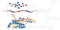
The receptor-binding domain, or RBD, is a crucial portion of a virus that allows it to latch to human cell receptors and therefore gain entry into cells and induce infection, according to researchers.
Moradi’s team’s models indicate the importance of the receptor-binding domain, but they also suggest that other regions, such as the N-terminal domain, may be important in the differences in SARS-CoV-1 and -2 spike protein binding behavior.
The N-terminal domain of a protein is a domain that is found at the beginning of the polypeptide chain, as opposed to the C-terminus, which is at the conclusion.
The role of the N-terminal region of SARS-CoV-1 and -2 spike proteins remains unknown, despite its proximity to the receptor-binding domain and the fact that it is known to be targeted by some antibodies. The research team led by Moradi is the first to discover evidence of a possible contact between the N-terminal domain and the receptor-binding domain.
“Our study sheds light on the conformational dynamics of the SARS-CoV-1 and SARS-CoV-2 spike proteins,” Moradi said. “Differences in the dynamic behavior of these spike proteins almost certainly contribute to differences in transmissibility and infectivity.”
The researchers’ study, “Prefusion Spike Protein Conformational Changes Are Slower in SARS-CoV-2 than SARS-Cov-1,” was published in Journal of Biological Chemistry.