Parotid Gland
Definition
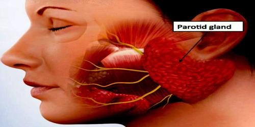
Parotid gland is either of the pair of salivary glands situated below and in front of each ear. In humans, the two parotid glands are present on either side of the mouth and in front of both ears. They are the largest of the salivary glands.
Approximately 20% of saliva is produced by parotid glands. This saliva is serous, that is, more liquid and fluid.
Certain diseases or the intake of substances like alcohol or certain drugs can affect the sympathetic nervous system (SNS), producing vasoconstriction of the parotid glands and reducing the secretion of saliva. This is the main reason why alcohol abuse is directly related to the lack of saliva.
Parotid glands produce a type of saliva that is “serous” which means it’s more watery and thin. It is has the protein Amylase that helps begin the process of starch digestion. While we are not eating, the parotid glands each contribute to 10% of saliva in the mouth, but when stimulated by eating the saliva each parotid gland produces accounts for 25% of the saliva in the mouth.
Structure and Functions of Parotide Gland
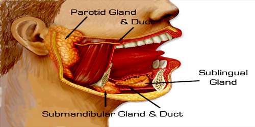
The parotid glands are located in front and beneath the ear. A duct, called Stensen’s duct, drains saliva from the parotid gland into the mouth, at the area of the upper cheeks. The submandibular glands are found on both sides, just under and deep to the jaw, towards the back of the mouth. This gland produces roughly 70% of the saliva in our mouth. The submandibular duct, called Warhtin’s duct, enter the floor of the mouth under the the front of the tongue. Sublingual glands, meanwhile, reside beneath the tongue, and supply saliva to the floor of the mouth as well. There are many (between 600 to 1,000) tiny glands called minor salivary glands. These glands are 1-2 mm in diameter and coat all the mucousal surfaces or lining of our mouth and throat.
The gland has four surfaces superficial or lateral, superior, anteromedial, and posteromedial. The gland has three borders anterior, medial, and posterior. The parotid gland has two ends superior end in the form of small superior surface and an inferior end (apex).
A number of different structures pass through the gland. From lateral to medial, these are:
- Facial nerve
- Retromandibular vein
- External carotid artery
- Superficial temporal artery
- Branches of the great auricular nerve
- Maxillary artery
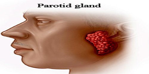
The function of the parotid gland and other two major salivary glands is to produce and secrete saliva, a substance that helps break food down so it can be digested properly. There are many different types of cells that make up the small little parts of the gland that produce saliva and secrete it. Because of the variety of cell types, there are many different types of tumors and cancers that can develop in the parotid gland.
The salivary glands are constantly working, and can be affected by many medical conditions, medications, and even not drinking enough water. Infections and inflammation of the gland can cause it to swell up and become painful. Obstruction of the ducts, which can happen because of salivary stones or narrowing of the duct from infection, can cause the saliva to back up into the gland and lead to it to swelling up as well.
Reference: