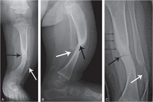
An X-ray is a quick and painless procedure that generates images of the inside of the body. It is a very effective method of inspecting bones and can be used to detect a variety of conditions. X-rays are typically performed by trained specialists known as radiographers in hospital X-ray departments, but they can also be performed by other healthcare professionals such as dentists.
At BESSY II, a team of medical researchers examined damage in bone samples from fish and mammals using focused high-energy X-rays. The scientists were able to document the destruction of collagen fibers caused by electrons emitted by mineral crystals using a combination of microscopy techniques. They conclude that X-ray methods may have an impact on bone samples when measured for an extended period of time.
Because it has long been known that X-rays damage living tissue beyond a certain dose, there are clear medical indications for X-rays to keep radiation exposure to a minimum. Researchers rely on increasingly powerful X-ray sources in basic research on the properties and characteristics of mineralized tissue samples such as bone.
We examined the samples under Second-Harmonic Generation laser-scanning microscopy for the imaging of the protein fibers. The high-energy photons from the X-ray light trigger a cascade of electron excitations. Ionization of calcium and phosphorus in the mineral then damages proteins like collagen in bone.
Katrein Sauer
Bones from fish and mammals
“Until now, the motto has actually been: more flux and higher energy is better, because you can achieve greater depth of field and higher resolution with more intense X-rays,” says Dr. Paul Zaslansky from Charité-Universitätsmedizin. Zaslansky and his team have now analyzed bone samples from fish and mammals at the MySpot beamline at BESSY II.
BESSY II generates a well characterized broad range of X-rays, precisely focused in an intermediate energy range which allows insights into the finest structures and even chemical and physical processes in materials. “Thanks to sensitive detectors and rather mild irradiation conditions in BESSY II as compared with harder X-ray synchrotron sources, we were able to demonstrate on our various bone samples that collagen fibers become damaged by the irradiation absorption in the mineral nanocrystals,” Zaslansky summarises the results of the study.
Imaging the protein fibers
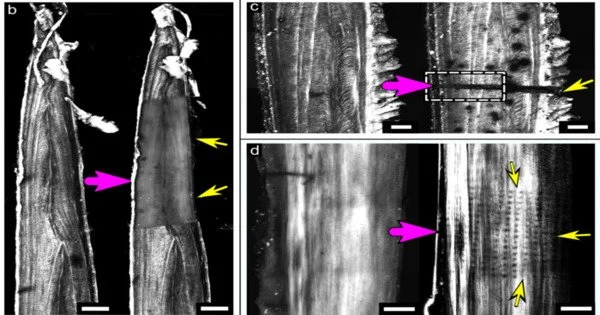
“We examined the samples under Second-Harmonic Generation laser-scanning microscopy for the imaging of the protein fibers” explains first author Katrein Sauer, who is doing her doctorate in Zaslansky’s team. Together with HZB expert Dr. Ivo Zizak, she irradiated bone samples from pike fish, pigs, cattle and mice with precisely calibrated X-ray light.
Trail of destruction
The beams left a trail of destruction that is clearly visible in the confocal and electron microscopy images. “The high-energy photons from the X-ray light trigger a cascade of electron excitations. Ionization of calcium and phosphorus in the mineral then damages proteins like collagen in bone,” Sauer says. Break-down of collagen increases with the duration of the irradiation, but also shows up even with short irradiation at high flux.
Minimal doses for research on living materials
“X-ray methods are considered non-destructive in materials research, but at least for research on bone tissues this is not true,” says Zaslansky. “We have to be more careful in basic medical research that we don’t damage the very structures we actually want to analyse.” So, as everywhere in medicine, and even when there are no living tissues and DNA to damage, it comes down to using a minimal dose to get the insights that reflect the material condition without causing damage.
Note:
The X-rays produced at BESSY II are about ten thousand times more intense than X-rays used for medical examinations (for X-rays of a broken leg, the German Federal Office for Radiation Protection gives a dose of 0.01 millisievert). X-ray methods are extremely useful for medical examinations.