Cancer treatments can be delivered to tumors in high dosages using nanoparticles while avoiding the negative side effects associated with chemotherapy. Only a small number of nanoparticle-based cancer medications have, however, received FDA approval.
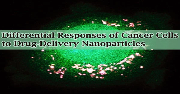
Researchers from MIT and the Broad Institute of MIT and Harvard have just published a study that may help to overcome some challenges facing the creation of medications based on nanoparticles. Thousands of biological characteristics determine whether cancer cells take up various forms of nanoparticles, according to the team’s examination of the interactions between 35 distinct types of nanoparticles and roughly 500 types of cancer cells.
The results may enable scientists to develop novel medication delivery particles that better target particular cancer kinds or better mimic the biological properties of specific cancer cell types.
“We are excited by our findings because it is really just the beginning we can use this approach to map out what types of nanoparticles are best to target certain cell types, from cancer to immune cells and other kinds of healthy and diseased organ cells. We are learning how surface chemistry and other material properties play a role in targeting,” says Paula Hammond, an MIT Institute Professor, head of the Department of Chemical Engineering, and a member of MIT’s Koch Institute for Integrative Cancer Research.
Hammond is the senior author of the new study, which appears in Science. The paper’s lead authors are Natalie Boehnke, an MIT postdoc who will soon join the faculty at the University of Minnesota, and Joelle Straehla, the Charles W. and Jennifer C. Johnson Clinical Investigator at the Koch Institute, an instructor at Harvard Medical School, and a pediatric oncologist at Dana-Farber Cancer Institute.
We are excited by our findings because it is really just the beginning we can use this approach to map out what types of nanoparticles are best to target certain cell types, from cancer to immune cells and other kinds of healthy and diseased organ cells. We are learning how surface chemistry and other material properties play a role in targeting.
Paula Hammond
Cell-particle interactions
Several other kinds of drug delivery nanoparticles have been created in the past by Hammond’s lab. Different cancer cell types frequently react differently to the same nanoparticles, according to studies conducted in her lab and elsewhere.
This phenomena was also noted by Boehnke, who was researching ovarian cancer at the time she joined Hammond’s lab, and Straehla, who was researching brain cancer. The researchers proposed that the diversity in the reactions of the cells may be caused by biological variations between cells.
They chose to do a large-scale investigation in which they could observe a great number of various cells interacting with a variety of nanoparticles in order to determine what those distinctions would be.
The PRISM platform, developed by the Broad Institute to enable researchers to swiftly screen thousands of medicines on hundreds of different cancer types simultaneously, was news to Straehla.
The team chose to try to modify that platform to evaluate cell-nanoparticle interactions rather than cell-drug interactions with the help of Angela Koehler, an associate professor of biological engineering at MIT.
“Using this approach, we can start thinking about whether there is something about a cell’s genotypic signature that predicts how many nanoparticles it will take up,” Boehnke says.
The researchers used 488 cancer cell lines from 22 different tissue origins for their screening. With the help of a distinctive DNA sequence, each type of cell is “barcoded,” enabling later identification by researchers. Extensive datasets on the gene expression profiles and other biological traits of each cell type are also accessible.
On the nanoparticle side, the researchers produced 35 particles, each with a core formed of either liposomes (particles made from many fatty molecules called lipids), PLGA, or polystyrene.
A variety of defense or targeting molecules, such as polymers like polyethylene glycol, antibodies, and polysaccharides, were also coated onto the particles by the researchers. This gave them the opportunity to research the effects of the particles’ surface chemistry and core composition.
The PRISM lab’s director, Jennifer Roth, and other Broad Institute researchers helped the researchers expose pools of hundreds of distinct cell types to one of 35 different nanoparticles.
Because each nanoparticle was fluorescently marked, the researchers were able to sort the cells according to how much fluorescence they released after being exposed for either four or 24 hours.
These measures were used to determine each cell line’s affinity for each nanoparticle, which was then converted into a score. The scores and all other available biological information for each cell line were then analyzed by the researchers using machine learning methods.
Numerous characteristics, or biomarkers, connected to an affinity for various kinds of nanoparticles were discovered through this investigation. The cellular machinery required to bind particles, transport them inside of cells, or process them is encoded by several of these markers, which were genes. Many of these genes were brand-new, although others of them were already known to be involved in the trafficking of nanoparticles.
“We found some markers that we expected, and we also found much more that has really been unexplored. We’re hoping that other people can use this dataset to help expand their view of how nanoparticles and cells interact,” Straehla says.
Particle uptake
The protein SLC46A3 was chosen as one of the biomarkers by the researchers for additional investigation. High quantities of this protein were associated with a very low uptake of lipid-based nanoparticles, according to the PRISM screen.
The same link was discovered when the researchers investigated these particles in melanoma mice models. According to the results, this biomarker may be utilized by medical professionals to identify patients whose tumors are more likely to respond to treatments including nanoparticles.
Researchers are currently working to determine the mechanism by which SLC46A3 controls the absorption of nanoparticles. They might be able to make tumors more responsive to medications delivered by lipid nanoparticles if they can find novel strategies to lower cellular amounts of this protein.
Additionally, the researchers are attempting to learn more about some of the additional biomarkers they discovered. Numerous additional types of nanoparticles that the researchers did not examine in this study could also be investigated using this screening method.
“The sky is the limit in terms of what other undiscovered biomarkers are out there that we just haven’t captured because we haven’t screened them,” Boehnke says. “Hopefully it’s an inspiration for others to start looking at their nanoparticle systems in a similar manner.”
The research was funded, in part, by SPARC funding to the Broad Institute, the Marble Center for Cancer Nanomedicine at the Koch Institute, and the Koch Institute Support (core) Grant from the National Cancer Institute.