Researchers have created a novel technique for quickly and easily producing different needle-shaped laser beams. Optical coherence tomography (OCT), a noninvasive imaging technique utilized for scientific research and numerous clinical diagnosis, can be improved with the use of these long, narrow beams.
“Needle-shaped laser beams can effectively extend the depth-of-focus of an OCT system, improving the lateral resolution, signal-to-noise ratio, contrast and image quality over a long depth range,” said research team leader Adam de la Zerda from Stanford University School of Medicine. “However, before now, implementing a specific needle-shaped beam has been difficult due to the lack of a common, flexible generation method.”
The researchers present their new platform for producing needle-shaped beams with various lengths and diameters in Optica, a journal published by Optica Publishing Group that focuses on high-impact research. It can be used to produce a variety of beams, including those with extraordinarily long depths of field and those that are narrower than the diffraction limit of light, for instance.
The needle-shaped beams generated with this method could benefit a variety of OCT applications. For instance, adopting a long, narrow beam could enable high-resolution OCT imaging of the retina without the need for dynamic focusing, speeding up the procedure and improving patient comfort. Additionally, it might increase the depth-of-focus for OCT endoscopy, which would increase the precision of the diagnostic.
“The rapid high-resolution imaging ability of needle-shaped beams can also get rid of adverse effects that occur due to human movements during image acquisition,” said the paper’s first author Jingjing Zhao. “This can help to pinpoint melanoma and other skin problems using OCT.”
A flexible solution
OCT has an axial resolution that remains constant throughout the imaging depth, making it a non-invasive imaging method. The light source determines its axial resolution, which has an extremely shallow depth of focus.
Needle-shaped beams can be used to improve the resolution of all microscopy systems, including particle manipulation with optical tweezers, materials processing, confocal microscopy, multiphoton microscopy, photolithography and photoacoustic tomography. Our model can also be applied to electromagnetic waves for terahertz imaging and even the mechanical waves used in ultrasound imaging.
Jingjing Zhao
In order to solve this problem, OCT devices are frequently designed with the ability to adjust the focus along the depth to take crisp pictures of an entire region of interest. However, this dynamic focussing can slow down imaging and is less effective in situations when the sample isn’t stationary.
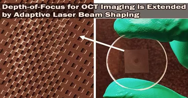
OCT frequently employs an objective lens that produces a single focal point with a single, condensed depth of field. The researchers employed a phase mask, a diffractive optical component that uses microstructures to produce different light patterns and produces several focal points along the axial direction, to improve the depth of focus.
To produce a new focus that is distinct from the original one, they constructed the phase mask using groups of pixels that were randomly dispersed and carefully patterned. In order to create densely spaced foci in the axial direction and create a needle-shaped beam with a deep depth of focus, the whole phase mask can then be used.
“Flexibility is the primary advantage of this new approach,” said Zhao. “Both the beam length and its diameter can be flexibly and accurately changed by modifying the locations of the foci and the phase difference between every two adjacent foci.”
This flexibility is made possible by a computational model the researchers created to precisely and quantitatively show the relationship between the beam qualities and the design parameters of the various foci. Based on the model’s calculations, they also created a high-performance production process to create diffractive optical components.
Selecting the right beam
The researchers designed beam shapes that could image a variety of materials to verify their approach. For example, to image individual cells within an entire layer of human epidermis, they created a needle-shaped beam with a diameter smaller than 2 microns (cellular resolution) and a length of at least 80 microns (epidermis thickness). Additionally, they were able to take high-resolution dynamic pictures of a beating heart in a drosophila larva, a crucial model organism for research into heart disease. To see the organ structure over a wide depth range, a beam that was 700 microns long and 8 microns in diameter was needed.
The team is currently attempting to improve the method by substituting a single flat metalens based on their model for the diffractive optical element and objective currently utilized to create a needle-shaped beam. For example, this metalens might be attached to a mouse’s cranium to allow real-time observation of the dynamics of the neuronal connections within the mouse brain.
The new work could also find applications beyond improving OCT.
“Needle-shaped beams can be used to improve the resolution of all microscopy systems, including particle manipulation with optical tweezers, materials processing, confocal microscopy, multiphoton microscopy, photolithography and photoacoustic tomography,” said Zhao. “Our model can also be applied to electromagnetic waves for terahertz imaging and even the mechanical waves used in ultrasound imaging.”