Basement Membrane
Definition
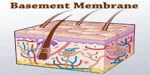
Basement membrane is a thin, delicate layer of connective tissue underlying the epithelium of many organs. It is also called basilemma. Basement membranes (BMs) are present in every tissue of the human body. All epithelium and endothelium is in direct association with BMs. BMs are a composite of several large glycoproteins and form an organized scaffold to provide structural support to the tissue and also offer functional input to modulate cellular function.
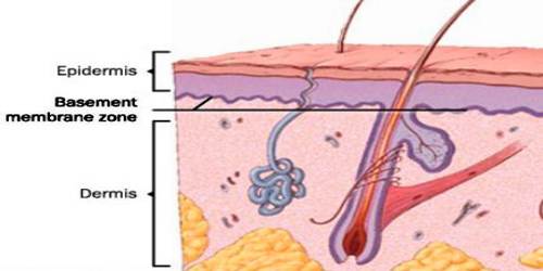
Some diseases result from a poorly functioning basement membrane. The cause can be genetic defects, injuries by the body’s own immune system, or other mechanisms. Genetic defects in the collagen fibers of the basement membrane cause Alport syndrome. Non-collagenous domain basement membrane collagen type IV is autoantigen (target antigen) of autoantibodies in the autoimmune disease Goodpasture’s syndrome.
A group of diseases stemming from improper function of basement membrane zone are united under the name epidermolysis bullosa.
The structure of BMs and their functional role in tissues are unique and unlike any other class of proteins in the human body. Increasing evidence suggests that BMs are unique signal input devices that likely fine tune cellular function. Additionally, the resulting endothelial and epithelial heterogeneity in human body is a direct contribution of cell-matrix interaction facilitated by the diverse compositions of BMs.
Structure and Functions of Basement Membrane
Basment membrane is an amorphous extracellular layer closely applied to the basal surface of epithelium and also investing muscle cells, fat cells, and Schwann cells; thought to be a selective filter and to serve both structural and morphogenetic functions; it is composed of three successive layers (lamina lucida, lamina densa, and lamina fibroreticularis), a matrix of collagen, and several glycoproteins.
Basement membrane contains a unique collagen, type IV collagen, which is formed of pro α1(IV) (Mr=185,000) and pro α2(IV) (Mr=170,000) chains. Type IV collagen has been localized to the basement membrane lamina densa, a nonfibrillar structure. Laminin is a large (Mr=1,000,000) noncollagenous glycoprotein with chains of 200,000 and 400,000 daltons. It has been localized to the basement membrane lamina lucida and functions to bind epithelial cells to the basement membrane. The basal lamina layer can further be divided into two layers. The clear layer closer to the epithelium is called the lamina lucida, while the dense layer closer to the connective tissue is called the lamina densa. The electron-dense lamina densa membrane is about 30–70 nanometers thick, and consists of an underlying network of reticular collagen IV fibrils which average 30 nanometers in diameter and 0.1–2 micrometers in thickness.
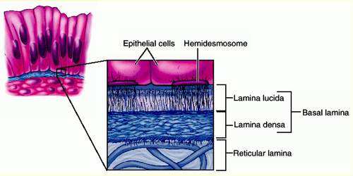
Its biological function may be to restrict the penetration of anionic macromolecules through the basement membrane. In contrast to the above-mentioned components which are found in all tissue basement membranes, bullous pemphigoid antigen is only found in certain basement membranes, mostly those of stratified squamous epithelia. Bullous pemphigoid antigen is a protein, synthesized by keratinocytes in culture, with disulfide-linked chains (Mr=220,000).
Basement membranes aren’t just found in the skin, though. They have important functions all over the body. Any place we find epithelium cells, which cover the inner and outer portions of glands, organs, and structural tissue, and endothelium tissue, which coats the inside of blood vessels, a basement membrane will be in between to hold the layers together. The most notable examples of basement membranes is the glomerular basement membrane of the kidney, by the fusion of the basal lamina from the endothelium of glomerular capillaries and the podocyte basal lamina, and between lung alveoli and pulmonary capillaries, by the fusion of the basal lamina of the lung alveoli and of the basal lamina of the lung capillaries, which is where oxygen and CO2 diffusion happens.
Reference:
