Aorta
Definition
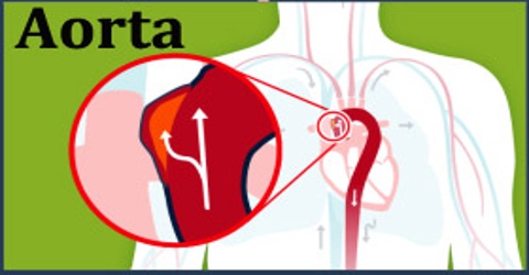
Aorta is the main artery of the circulatory system, arising from the left ventricle of the heart in mammals and birds and carrying blood with high levels of oxygen to all the arteries of the body except those of the lungs. The word ‘Aorta’ stems from the Late Latin Latin: aorta from aortē (Greek: ἀορτή), from aeirō, “I lift, raise” (Greek: ἀείρω) This term was first applied by Aristotle when describing the aorta and describes accurately how it seems to be ‘suspended’ above the heart.
The aorta is traditionally divided into the ascending aorta, the aortic arch, and the descending aorta. The descending aorta is, in turn, subdivided into the thoracic aorta that descends within the chest and the abdominal aorta that descends within the belly. The function of the aorta has been documented in the Talmud, where it is noted as one of three major vessels entering or leaving the heart, and where perforation is linked to death.
The heart is the organ of the cardiovascular system that functions to circulate blood along pulmonary and systemic circuits. The aorta arises from the left ventricle of the heart, forms an arch, and then extends down to the abdomen where it branches off into two smaller arteries. Several arteries extend from the aorta to deliver blood to the various regions of the body.
Structure and Functions of Aorta
The aorta is a tube about a foot long and just over an inch in diameter. The aorta is divided into four sections:
- The ascending aorta rises up from the heart and is about 2 inches long. The coronary arteries branch off the ascending aorta to supply the heart with blood.
- The aortic arch curves over the heart, giving rise to branches that bring blood to the head, neck, and arms.
- The descending thoracic aorta travels down through the chest. Its small branches supply blood to the ribs and some chest structures.
- The abdominal aorta begins at the diaphragm, splitting to become the paired iliac arteries in the lower abdomen. Most of the major organs receive blood from branches of the abdominal aorta.
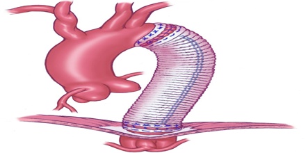
The walls of the aorta consist of three layers. They are the tunica adventitia, the tunica media, and the tunica intima. These layers are composed of connective tissue, as well as elastic fibers. These fibers allow the aorta to stretch to prevent over-expansion due to the pressure that is exerted on the walls by blood flow.
Variations may occur in the location of the aorta, and the way in which arteries branch off the aorta. The aorta, normally on the left side of the body, may be found on the right in dextrocardia, in which the heart is found on the right, or situs inversus, in which the location of all organs are flipped.
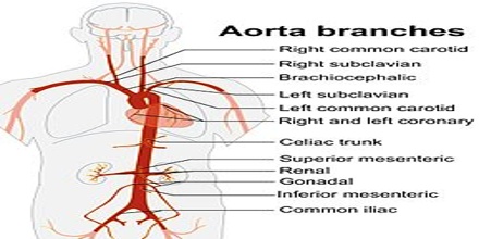
The aorta supplies all of the systemic circulation, which means that the entire body, except for the respiratory zone of the lung, receives its blood from the aorta. Broadly speaking, branches from the ascending aorta supply the heart; branches from the aortic arch supply the head, neck and arms; branches from the thoracic descending aorta supply the chest excluding the heart and the respiratory zone of the lung; and branches from the abdominal aorta supply the abdomen. The pelvis and legs get their blood from the common iliac arteries. The difference between aortic and right atrial pressure accounts for blood flow in the circulation. When the left ventricle contracts to force blood into the aorta, the aorta expands.
Reference: webmd.com, thoughtco.com, dictionary.com, wikipedia.