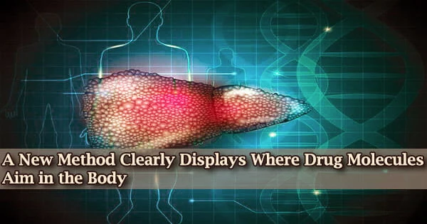
Researchers at Scripps Research have developed a method to more precisely and across a variety of tissues scan the locations in the body where medications attach to their intended targets. The novel approach might be regularly used in drug development.
The new technology, dubbed CATCH, is explained in a study published in Cell on April 27, 2022. It involves attaching fluorescent tags to medication molecules and using chemical methods to enhance the fluorescent signal.
Chemical rings, chains, and functional groups are frequently found in pharmacological molecules. Chemical rings play a significant role in medication structures. The strategy was proven by the researchers using a variety of experimental medications, demonstrating where the drug molecules found their targets even within individual cells.
“This method ultimately should allow us, for the first time, to see relatively easily why one drug is more potent than another, or why one has a particular side effect while another one doesn’t,” says study senior author Li Ye, Ph.D., assistant professor of neuroscience at Scripps Research and The Abide-Vividion Chair in Chemistry and Chemical Biology.
Zhengyuan Pang, the study’s first author, is a graduate student in the Ye lab. The Gilula Chair of Chemical Biology at Scripps Research and Ben Cravatt’s lab worked closely together on the study as well.
“The unique environment at Scripps Research, where biologists routinely work together with chemists, is what made the development of this technique possible,” Ye says.
A fundamental component of medication research is understanding where drug molecules bind their targets to exert their therapeutic effects and adverse effects. Drug-target interaction investigations, however, have typically used very sloppy techniques, like bulk assessments of drug-molecule concentration in whole organs.
The unique environment at Scripps Research, where biologists routinely work together with chemists, is what made the development of this technique possible.
Li Ye
Small chemical handles are inserted into medication molecules as part of the CATCH technique. The insertion of fluorescent tags is possible after the medication molecules have bonded to their targets thanks to these specific chemical handles, which do not interact with any other substances in the body.
Ye and his team paired the tagging process with a method that renders tissue relatively transparent, in part because human or animal tissue has a tendency to disperse and obscure the light from these fluorescent tags.
For “covalent medicines,” which bind to their targets with stable chemical connections known as covalent bonds, the researchers optimized and assessed their strategy in this initial investigation. It is crucial to ensure that these medications are reaching their targeted targets because the binding is irreversible.
The researchers first examined a number of covalent inhibitors of the brain’s fatty acid amide hydrolase (FAAH) enzyme. FAAH inhibitors are being researched as therapies for pain and mood disorders because they increase amounts of cannabis compounds, such as the “bliss molecule” anandamide.
The researchers at the single-cell level imaged large areas of mouse brain tissue, allowing them to identify the precise locations of these inhibitors’ targets and distinguish between their various patterns of target engagement.
In one experiment, they demonstrated that an experimental FAAH inhibitor called BIA-10-2474 engages unidentified targets in the midbrain of mice even when the mice lack the FAAH enzyme, providing a clue to the cause of the inhibitor’s toxicity. In a clinical trial in France in 2016, the inhibitor caused one death and multiple injuries.
In other experiments showcasing the novel technique’s unmatched accuracy and adaptability, the researchers revealed that they could combine drug-target imaging with several fluorescent-tagging techniques to identify the cell types to which a drug binds. Additionally, they were able to identify drug-target contact sites in several neuronal regions.
Finally, scientists were able to observe how slightly changing drug doses frequently had a noticeable impact on the level of target engagement in various brain regions. Ye underlines that the proof-of-principle work is merely the beginning. In the future, maybe whole mice, he and his team hope to apply CATCH on thicker tissue samples.
They also intend to expand the scope of the fundamental strategy to include more widely used chemical probes and non-covalently bound pharmaceuticals. Overall, the new technology, according to Ye, is seen as a fundamental tool for both drug discovery and fundamental biology.
“In situ Identification of Cellular Drug Targets in Mammalian Tissue,” was co-authored by Zhengyuan Pang, Michael Schafroth, Daisuke Ogasawara, Yu Wang, Victoria Nudell, Neeraj Lal, Dong Yang, Kristina Wang, Dylan Herbst, Jacquelyn Ha, Carlos Guijas, Jacqueline Blankman, Benjamin Cravatt and Li Ye all of Scripps Research during the study.
The National Institutes of Health (DP2DK128800, DK114165, DK124731, DA033760), the Whitehall Foundation, the Baxter Foundation, and the Dana Foundation funded the study in part.