Stem-cell produced heart tissues have a bright future for therapeutic uses to treat cardiac illness, according to research in animal models. Scientists must first accurately grasp at the cellular and molecular levels which conditions are required for implanted stem-cell derived cardiac cells to develop and integrate in three dimensions inside surrounding tissue before such therapies are viable and safe for use in humans.
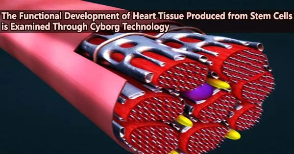
New findings from the Harvard John A. Paulson School of Engineering and Applied Sciences (SEAS) make it possible for the first time to monitor the functional development and maturation of cardiomyocytes the cells responsible for regulating the heartbeat through synchronized electrical signals on the single-cell level using tissue-embedded nanoelectronic devices. The devices, which are flexible, stretchable, and can seamlessly integrate with living cells to create “cyborgs”, are reported in a Science Advances paper.
“These mesh-like nanoelectronics, designed to stretch and move with growing tissue, can continuously capture long-term activity within individual stem-cell derived cardiomyocytes of interest,” says Jia Liu, co-senior author on the paper, who is an assistant professor of bioengineering at SEAS, where he leads a lab dedicated to bioelectronics.
In order to bridge the gap between living tissue and electronics, Liu’s team has developed a number of flexible, minimally invasive mesh-like nanoelectronic sensors that can be embedded in natural tissues without interfering with cellular growth or function.
“Nature showed us the solution to monitoring tissue in 3D,” Liu says. “We were inspired by the way neural tubes fold during development, stretching as cells migrate and take shape into tissue volume.”
If we have both nanoelectronic sensors and stimulators, we can monitor electrical activity and use feedback to pace implanted tissues into the same frequency as surrounding tissues. This approach could be adapted to so many other types of stem-cell-derived tissues, such as neuronal tissues and pancreatic organoids.
Jia Liu
In order to test the idea of using a mesh-like nanoelectronic structure, his team built their first cyborg organoid in 2019. They have already shown that these flexible nanoelectronics can be transplanted into living mice without endangering the functionality of the cells around them.
In their latest study, Liu’s lab joined forces with Richard Lee and his team at Harvard Stem Cell Institute and used the nanoelectronics to monitor electrical activity of stem-cell derived cardiomyocytes.
To do so, the researchers cultured cells onto a sheet made of commercially available cellular matrix known as “Matrigel” and the mesh-like nanoelectronic sensor (which contains a flexible grid of microelectrodes).
The researchers saw that the sheet easily stretched and accommodated the stem-cell generated tissues as they proliferated and expanded in 3D as the cells matured and formed into a tiny organoid structure.
By employing these methods in in vitro research, the team learned that the endothelial cells, which control blood flow between blood vessels and surrounding tissues, play a previously underappreciated but significant role in the quick and effective maturation of stem-cell derived cardiomyocytes.
When cultured together in a 3D cardiac tissue matrix, cardiomyocytes underwent “extraordinary electrical maturation” in the presence of endothelial cells.
The scientists saw a direct correlation between proximity to endothelial cells and the development of the organoids throughout the duration of seven weeks of observation. In addition to maturing more quickly than cardiomyocytes that were cultivated at a distance from endothelial cells, the closer-located cardiomyocytes also exhibited electrical properties that are typical of heart tissue in a healthy state.
The new understanding is a major step forward for creating heart tissues from stem cells. It is challenging to create and transplant cardiomyocytes derived from stem cells that can beat in unison with surrounding heart tissue for prolonged periods of time, according to experimental preclinical study on animals with hearts that resemble those of humans. The electrical misfire that results from immature cardiomyocytes being implanted into an animal’s heart might result in dangerously irregular heartbeats.
That’s why the discovery that co-culturing stem-cell-derived cardiomyocytes with endothelial cells can create more functionally mature cardiomyocytes is so significant.
The team’s new paper also describes the use of a novel machine-learning-based analysis to decipher the electrical activity recorded by the tissue-embedded nanoelectronic devices, allowing for continuous monitoring of the electrical waves produced by the target cardiomyocytes as they mature and improving knowledge of how the tissue microenvironment affects electrical stability.
According to Liu, the nanoelectronic devices and machine-learning-based analysis represent new platform technologies for monitoring and managing stem-cell derived tissue implants, allowing researchers to cultivate cyborgs made of both living tissues and electronics that can be controlled with great precision.
He even imagines a future in which these cyborgs could be used in cardiac tissues to detect abnormal electrical activity in cardiomyocytes and deliver highly targeted voltage, acting as a nanoscale pacemaker, to help correct implanted cells and ensure they continue to beat in rhythm with the rest of the heart.
“If we have both nanoelectronic sensors and stimulators, we can monitor electrical activity and use feedback to pace implanted tissues into the same frequency as surrounding tissues,” Liu says. “This approach could be adapted to so many other types of stem-cell-derived tissues, such as neuronal tissues and pancreatic organoids.”
Additionally, he claims that this nanoelectronics platform technique might be applied to drug screening, offering continuous, single-cell-level study of how tissues react to various medications and treatments.
