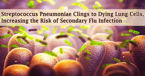
A flu virus infection is difficult enough, but when Streptococcus pneumonia is added to the mix, it can be fatal. Researchers have discovered a new virulence mechanism for a surface protein on the pneumonia-causing bacteria S. pneumoniae, which could explain why this dual infection is so severe.
This breakthrough comes more than three decades after the pneumococcal surface protein A, or PspA, was discovered. This novel technique had previously gone unnoticed because it only allows germs to stick to dead or dying lung epithelial cells, not living ones.
Previously, researchers looked for bacterial adhesins that assist infection in healthy lung cell monolayers. Virus-induced lung cell death during the flu has been found to pave the route for S. pneumonia to attach to the airway, exacerbating the illness and pneumonia.
Carlos Orihuela, Ph.D., and David Briles, Ph.D., professor and professor emeritus in the University of Alabama at Birmingham Department of Microbiology, conducted the study, which was published in the journal Cell Reports.
Orihuela and Briles claim that these findings add to the understanding of how an influenza A flu virus infection followed by S. pneumoniae superinfection results in severe pneumonia and a high fatality rate. The technique could possibly lead to advancements in disease treatment and vaccination.
Banked lung samples from the 1918 Spanish influenza pandemic, which killed 40 million to 50 million people, provide a historical example of the deadly synergy of flu infection followed by S. pneumoniae superinfection. The vast majority of these samples showed co-infection or secondary infection with S. pneumonia.
Our findings support the targeting of regions of PspA for therapeutic and vaccine development against influenza A/Streptococcus pneumoniae superinfections. Importantly, and despite more than 30 years since its discovery, PspA was not previously shown to function as an adhesin. Thus, our finding of PspA’s role in adherence substantially advances our knowledge on the interactions of S. pneumoniae with its host.
Carlos Orihuela
The PspA research at UAB started with some perplexing results from experimental influenza A lung infections in mice, followed by either wild-type S. pneumoniae with the intact PspA gene or a mutant S. pneumoniae lacking PspA.
In comparison to lungs infected with the mutant, lung homogenates from mice infected with the wild-type exhibited a substantially larger amount of S. pneumonia bacteria. When researchers cleansed the insides of the lungs and collected the bronchoalveolar lavage fluid, they found that the wild-type S. pneumonia and the mutant had identical amounts of bacteria.
“This unexpected result was interpreted to mean that wild-type S. pneumoniae were more resistant to dislodgement than S. pneumonia with a pspA gene deletion, and it served as rationale for further experimentation,” Orihuela said.
The researchers were able to prove that, in addition to its other known virulence mechanisms, PspA acts as an adhesin to dying host cells based on this hint. The molecular mechanism of bacterial adhesion was also described by the researchers.
Lung epithelial cells die from both influenza A infection and the production of the S. pneumoniae toxin pneumolysin. Phosphatidylserine residues in dying cells are flipped to the outer cell membrane, where they bind the host enzyme GAPDH (glyceraldehyde-3-phosphate dehydrogenase).
In turn, the S. pneumoniae PspA protein on the bacteria’s surface binds to GAPDH. S. pneumoniae localisation in the lower airway was boosted by PspA-GAPDH-mediated binding to lung cells, which was aided by pneumolysin exposure or co-infection with influenza A virus.
GAPDH binding was found to need a 52-amino-acid section of the PspA protein from amino acids 230 to 281 in tests utilizing fragments of the protein. Through binding competition, instilling one of those binding fragments into the lungs of influenza-infected mice lowered the severity of S. pneumoniae superinfection.
“Our findings support the targeting of regions of PspA for therapeutic and vaccine development against influenza A/Streptococcus pneumoniae superinfections,” Orihuela said.
“Importantly, and despite more than 30 years since its discovery, PspA was not previously shown to function as an adhesin. Thus, our finding of PspA’s role in adherence substantially advances our knowledge on the interactions of S. pneumoniae with its host.”