Doctors and researchers must be able to peer into bodies in order to properly identify and cure illnesses. Since the invention of the first x-ray, medical imaging techniques have advanced significantly, but the majority of them are still too crude to count or identify specific cell types in deep tissues of the body.
Quantum dots can do that, according to new research in mice from the University of Illinois.
“Quantum dots can measure things in the body that are very, very dynamic and complicated and that we can’t see currently. They give us the ability to count cells, detect their exact locations, and observe changes over time. I think it is really a huge advance,” says Andrew Smith, professor in the Department of Bioengineering at U of I and co-author on the ACS Nano study.
Few hundred atom-sized quantum dots are lab-grown nanoparticles with unique optical properties that can be seen via fluorescence imaging, tomography (like PET/CT scanners), and standard microscopy. Bioengineers like Smith can make them glow in a certain color and produce infrared light depending on their size and makeup.
“Emitting light in the infrared is rare. Very little light is emitted by tissues in the infrared, so if you put them in the body, they appear very bright. We can see deeply into the body and can more accurately measure things than we could using technology in the visible range,” Smith says.
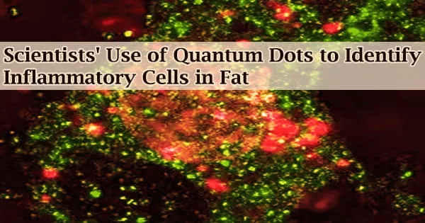
In the ACS Nano study, Smith and colleagues let quantum dots loose on macrophages.
Macrophages get to work when our systems need to consume infections or remove cellular trash. One of its tasks is to start an inflammatory response that renders the environment hostile to dangerous microorganisms.
Quantum dots can measure things in the body that are very, very dynamic and complicated and that we can’t see currently. They give us the ability to count cells, detect their exact locations, and observe changes over time. I think it is really a huge advance.
Professor Andrew Smith
But occasionally they perform their duty too competently. Chronic inflammation brought on by macrophage activity can cause diabetes, cardiovascular problems, malignancies, and more, depending on the tissue in which it occurs. The U of I researchers was especially curious about macrophages in adipose tissue, also known as fat.
“With weight gain and obesity, macrophage numbers are known to increase in adipose tissue and tend to shift towards an inflammatory phenotype, which contributes to the development of insulin resistance and metabolic syndrome. The number and location of macrophages in adipose tissue are poorly described, especially in vivo,” says Kelly Swanson, Kraft Heinz Company Endowed Professor in Human Nutrition in the Department of Animal Sciences at U of I and study co-author.
“The quantum dots our group developed allow for better quantification and characterization of the cells present in adipose tissue and their spatial distribution,” he adds.
Dextran is a sugar molecule that also targets macrophages in adipose tissue, and the team made quantum dots coated with it. They gave these quantum dots to obese mice as a proof-of-concept and compared the imaging outcomes to dextran alone, the industry standard for imaging macrophages.
Using simple optical methods as well as all imaging platforms, quantum dots outperformed dextran alone.
“Quantum dots put out a huge amount of light, giving us the ability to measure specific cell types to a greater degree and identify where they are,” Smith says. “That degree of light output allows the use of optical techniques, which are much more accessible than other imaging technologies. Compared with MRI and PET scanners, they’re cheap instruments that can be put into a small clinic. Everybody could have one.”
Swanson envisions a time when a straightforward optical tool, such as ultrasonography, may be used to non-invasively identify and monitor inflammatory macrophages in patients who are overweight even if quantum dots have not yet been employed in humans.
“There could be a device like an ultrasound where you scan somebody, and even if a patient’s weight hasn’t changed, a doctor could tell if the cell types are changing. More inflammatory cells could predict insulin resistance and other issues,” he says. “That’s why I’m interested in it, for its diagnostic properties.”
Because they are frequently manufactured with toxic metals like cadmium and mercury and because researchers are still trying to understand how they are metabolized and eliminated from the body, quantum dots aren’t used in people.
While Smith and his team are developing quantum dots composed of safer materials, they are still a crucial research tool in the interim. For instance, their prolonged circulation period, which in the present study was nine times longer than that of dextran, may allow diagnosticians to move beyond a single point in time.
“There’s a huge level of variability of macrophages even across a day. Adipose tissue may have a very high number in the middle of the day, and then it drops way down,” Smith says. “In animal studies, we can sacrifice animals at the start and end of a day to study the trend, but with quantum dots, we might not have to do that. You could track one animal over time to see its progression. Quantum dots offer a huge amount of value in animal studies. So even if quantum dots don’t make it to humans, if we never find a way to make them non-toxic, the value is still really great.”
A funding from the National Institutes of Health was recently given to Smith, Swanson, and other researchers to broaden their use of quantum dots to target numerous different cell types.
At the University of Illinois Urbana-Champaign, the Department of Animal Sciences is a part of the College of Agricultural, Consumer, and Environmental Sciences.