The biomedical research community is faced with a challenging task as a result of aging and age-related illnesses. It is crucial to comprehend how senescence is controlled in order to promote healthy aging and address illnesses linked to old age.
Rugang Zhang, Ph.D., the Christopher M. Davis Endowed Professor and program director of the Immunology, Microenvironment & Metastasis Program at The Wistar Institute, and his team described a novel ADAR1-SIRT1-p16INK4a axis in regulating cellular senescence and its potential implications in tissue aging in a study that was published today in Nature Cell Biology.
Proteins are called enzymes to aid in accelerating our bodies’ chemical reactions, or metabolism. Some compounds are created, while others are broken down. Enzymes are a part of all living things. Enzymes are created by our bodies spontaneously. However, food and manufactured goods both contain enzymes.
“Understanding the basic mechanism underlying tissue aging is challenging and cellular senescence offers an angle into the complex biology that drives tissue aging. These mechanistic insights gained by studying senescence regulation during tissue aging can, in turn, be used to promote healthy aging and combat age-associated disorders,” states Zhang.
The protein p16INK4a, whose expression both rises with tissue aging and promotes senescence, is essential to this search. Previous research demonstrated that the removal of cells that express the p16INK4a gene is sufficient to postpone age-related diseases.
Therefore, methods that stop the age-related increase in p16INK4a expression may have significant effects on the creation of intervention strategies that support healthy aging. The findings of the study team are centered on a protein called ADAR1. A specialized enzyme called ADAR1 that participates in RNA editing has recently been linked to senescence.
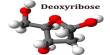
All living cells contain ribonucleic acid, or RNA, a nucleic acid with structural similarities to DNA (Deoxyribonucleic Acid). However, RNA is often single-stranded, unlike DNA. Instead of the deoxyribose present in DNA, the backbone of an RNA molecule is made up of alternating phosphate groups and sugar ribose.
Understanding the basic mechanism underlying tissue aging is challenging and cellular senescence offers an angle into the complex biology that drives tissue aging. These mechanistic insights gained by studying senescence regulation during tissue aging can, in turn, be used to promote healthy aging and combat age-associated disorders.
Rugang Zhang
Xue Hao, Ph.D., a postdoctoral researcher in the Zhang lab and the paper’s first author, explains that earlier independent studies in model organisms like fruit flies and worms showed that the depletion of the human ADAR1 equivalent in these organisms shortens lifespan and results in age-dependent changes like neurodegeneration.
The Wistar Institute’s ethos of intense cooperation also helps this story. In fact, prior research by Kazuko Nishikura, Ph.D., a pioneer in ADAR1 biology and professor in the Gene Expression & Regulation Program at Wistar’s Ellen and Ronald Caplan Cancer Center, demonstrated that stressed cells use ADAR1 as protection from apoptosis, or programmed cell death.
“As senescent cells are stressed cells and are resistant to apoptosis, the first question we set out to ask was whether ADAR1 is related to cellular senescence and secondly, how does it regulate senescence and what is its’ potential implication in tissue aging.” Hao explains.
First, the team looked at the expression of ADAR1 in vivo in several organs from young and old mice, as well as in vitro in human fibroblasts. Then, in order to prove that ADAR1 is an essential regulator of p16INK4a expression, they experimentally changed the expression of ADAR1 in several cell types in petri dishes and mouse tissues.
Surprisingly, the scientists found that SIRT1, another protein with a reputation for controlling both senescence and tissue aging, mediates the effect of ADAR1 depletion on p16INK4a expression. It’s interesting to note that this ADAR1 function is independent of its biological function in RNA editing.
Additionally, they discovered that senescence decreased the stability of SIRT1 mRNA, which in turn increased the translation of p16INK4a to induce senescence. This was due to the downregulation of ADAR1 by a process known as autophagy (the degradation and recycling of harmed or unnecessary cell components).
Hao elaborates, “Our study revealed a novel ADAR1-SIRT1- p16 INK4a axis that plays an important role in cellular senescence at the translational level, and this newly defined function of ADAR1 is independent of its RNA editing function.”
Zhang says, “Our study starts to reveal the missing link between ADAR1 and tissue aging through p16INK4a expression during senescence. In addition, these findings provided a scientific rational to explore whether this newly discovered mechanism can be leveraged for therapeutic development regarding age-associated disorders.”
“One of the ways to potentially restore ADAR1 expression as a means to suppress p16INK4a and senescence observed during tissue aging is by inhibiting autophagy.” Hao details. She adds about next research steps,
“Our study raises some interesting questions. For example, what is the relative contribution of this mechanism to p16INK4a expression during the aging of different tissues? In addition, it would be interesting to determine whether the intervention of this pathway can alleviate the age-associated disorders that are linked to p16INK4a expression in previous published animal models.”
Co-authors: Xue Hao, Yusuke Shiromoto, Masayuki Sakurai, Martina Towers, Qiang Zhang, Shuai Wu, Bin Tian, Andrew Kossenkov, Kazuko Nishikura, Pingyu Liu from The Wistar Institute; Aaron Havas, Peter D. Adams from Sanford Burnham Prebys Medical Discovery Institute; Lu Wang, Shelley Berger from the University of Pennsylvania.
Work supported by: This work was supported by US National Institutes of Health grants (R01CA160331 to R.Z., P01AG031862 to P.D.A., S.L.B, and R.Z., R01GM040536 and R01GM130716 to K.N., and R50CA211199 to A.V.K.). K.N. was supported by a grant from Emerson Collective. Support of Core Facilities was provided by Cancer Center Support Grant (CCSG) CA010815 to The Wistar Institute.