The idea of “spying” on a shape-shifting protein may sound like science fiction, yet investigating the dynamic behavior of proteins, especially those that undergo shape-shifting or conformational changes, is a legitimate and essential field of scientific study. Crystallography is being used by researchers to improve their understanding of protein shapeshifting. The knowledge can help prevent and treat diseases.
Proteins carry out biological processes in our bodies by interacting with metabolites or other proteins to fulfill tasks. To do this, protein molecules frequently shape-shift to enable specialized binding interactions required for sophisticated, precise chemical processes.
A deeper understanding of the shapes proteins assume might help researchers prevent or treat diseases, but existing approaches for disclosing these dynamic, three-dimensional structures provide scientists with limited information. To fill this knowledge gap, a team from the CUNY Graduate Center’s Advanced Science Research Center (CUNY ASRC) devised an experiment to see if imaging X-ray crystallography at elevated temperature versus elevated pressure yielded unique morphologies. The team’s findings appear in the journal Communications Biology.
Protein structures don’t sit still; they shift between several similar shapes much like a dancer. Unfortunately, existing approaches for viewing proteins only reveal one shape, or suggest the presence of multiple shapes without providing specific details.
Daniel Keedy
“Protein structures don’t sit still; they shift between several similar shapes much like a dancer,” said the study’s principal investigator Daniel Keedy, Ph.D., a professor with the CUNY ASRC’s Structural Biology Initiative and a chemistry and biochemistry professor at The City College of New York and the CUNY Graduate Center.
“Unfortunately, existing approaches for viewing proteins only reveal one shape or suggest the presence of multiple shapes without providing specific details. We wanted to see if different ways of poking at a protein could give us a more detailed view of how it shape-shifts.”
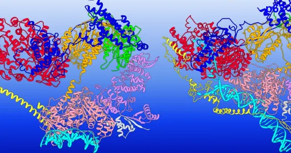
For their experiment, the team obtained crystals of STEP, also known as PTPN5 — a drug target protein for the treatment of several diseases, including Alzheimer’s — and agitated them using either high pressure (2,000 times the Earth’s atmospheric pressure) or high temperature (body temperature), both of which are very different from typical crystallography experiments at atmospheric pressure and cryogenic temperature (-280 F, -173 C). The researchers viewed the samples using X-ray crystallography and observed that high temperature and high pressure had different effects on the protein, revealing distinct shapes.
While high pressure is not a condition that proteins face within the body, Keedy believes the agitation method revealed multiple structural states of the protein that may be important to its activity in human cells.
“Having the ability to use perturbations such as heat and pressure to elucidate these different states could give drug developers tools for determining how they can trap a protein in a particular shape using a small-molecule drug to diminish its function,” Keedy said in a statement.