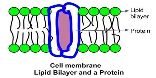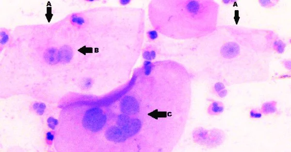
Nuclear atypia is characterized by the abnormal appearance of cell nuclei. It refers to changes in the size, shape, and appearance of cell nuclei within tissues that are abnormal. It is a cytopathology and histopathology term. Pleomorphic nuclei are common in atypical nuclei. The central components of cells that contain genetic material (DNA) are known as nuclei. Cell nuclei have a consistent and uniform appearance under normal conditions.
Atypia of the nucleus can be seen in reactive changes, pre-neoplastic changes, and malignancy. In most cases, severe nuclear atypia is considered a sign of cancer.
However, nuclear atypia can occur as a result of various conditions, including but not limited to:
- Cancer: Cancer cells often exhibit nuclear atypia. The nuclei in cancer cells may become larger, more irregular in shape, and may have abnormal chromatin patterns. These changes are indicative of uncontrolled cell growth and a loss of normal cellular regulation.
- Inflammation: Changes in cell nuclei can occur as a result of inflammatory conditions. Chronic inflammation can cause changes in cell structure and function, such as nuclear atypia.
- Infections: Changes in cell nuclei can also be caused by viral or bacterial infections. In response to the infection, the host cell may exhibit atypical nuclear features.
- Dysplasia: Dysplasia is defined as abnormal cell or tissue development. Dyplastic cells in the context of nuclear atypia may exhibit irregularities in nuclear size and shape, indicating a potential precursor to cancer.
Nuclear atypia detection is frequently an important aspect of histopathological examination, which involves examining tissue samples under a microscope. Pathologists examine these changes to determine the nature of a tissue, diagnose diseases, and determine the likelihood of malignancy.