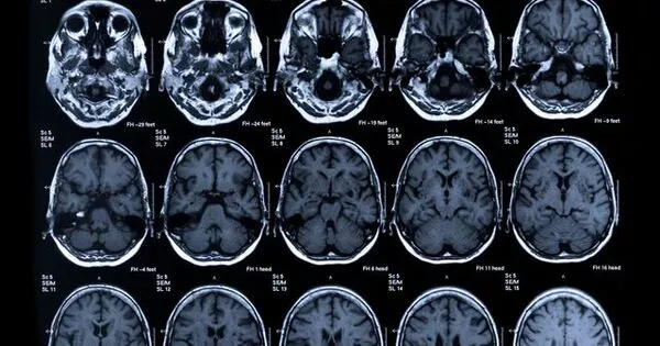
New technology, such as functional magnetic resonance imaging (fMRI) and optical imaging, has allowed for better visualization of the brain, particularly in the study of the “little brain” or cerebellum. These techniques have revealed previously unknown functions of the cerebellum, including its role in cognitive processes and movement coordination. Additionally, advances in imaging methods have allowed for more detailed visualization of the cerebellum’s intricate structure, aiding in the understanding of brain disorders and disease.
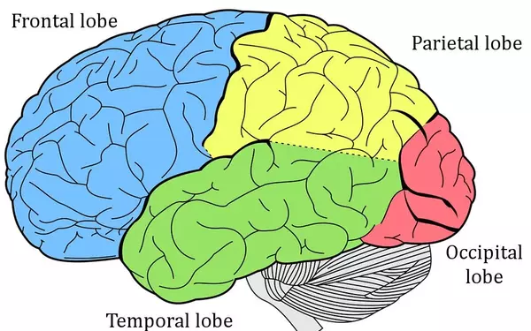
The cerebellum, or our little brain, is primarily in charge of our motor skills. In addition, structure is important for behavior and cognition. The cerebellum is a part of your brain that is located at the back of your head, just above and behind the point where your spinal cord connects to your brain. Although it only accounts for 10% of our brain’s volume, the cerebellum contains more brain cells than the rest of our brain and is thus an important area to properly map out.
In humans, the cerebellum is more folded than in other mammals. Because of the condensed layers, this makes it a difficult structure to fully visualize and thus study. High resolution imaging is required to properly image it. Only a small portion of the cerebellar anatomy could be visualized using current imaging techniques, leaving the remaining details out. As a result, the ingenious structure remains largely unknown.
We can do this specifically because we have a very high field magnet and also motion correction because people tend to move during the scans. We can now investigate the cerebellum’s cognitive role as well as its clinical implications.
Nikos Priovoulos
High resolution
Researchers at the Spinoza Center have developed a method to look at the cerebellum using a powerful 7-Tesla MRI scanner. By correcting for movements during, for example, the subjects’ breathing, the team succeeded in achieving a resolution of 1/5th of a millimetre. In addition, the researchers can use this data to reconstruct and digitally inflate the cerebellum, making the patterns and layers clearly visible. The cerebellum can also be viewed ‘live’ while performing various tasks.
New technology, such as advanced imaging techniques like magnetic resonance imaging (MRI) and positron emission tomography (PET) scans, can provide detailed visualizations of the brain and its structures. These technologies can help researchers and medical professionals better understand the brain and its functions, and aid in the diagnosis and treatment of brain-related disorders. Additionally, advancements in computer software and algorithms can enhance the resolution and accuracy of these images, providing even more detailed views of the brain.
‘We can do this specifically because we have a very high field magnet (which is expensive and difficult to build) and also motion correction because people tend to move during the scans,’ says researcher Nikos Priovoulos. We can now investigate the cerebellum’s cognitive role as well as its clinical implications. We hope that by using this technique, we will be able to look more closely at neurodegenerative disorders. This is the first time we have been able to see the human cerebellum in such detail.’
‘In multiple sclerosis (MS), the cerebellum plays an important role. MS patients have motor lesions, which means that the nerve cells that control movement have been damaged. Ex vivo studies, or experiments performed outside the body, have already revealed that degeneration of these cells occurs in MS. We are currently scanning MS patients and hope to look at other clinical groups as well. Based on previous research, we know that high resolution imaging in the cerebellum could help with MS. We don’t know what the long-term impact will be. Hopefully, it has some predictive value for these patients.’