Medulla Oblongata
Definition
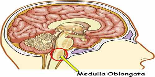
Medulla oblongata is the lowermost portion of the brainstem in humans and other mammals. It is important in the reflex control of involuntary processes, including respiration, heartbeat, and blood pressure. It is a cone-shaped neuronal mass responsible for autonomic (involuntary) functions ranging from vomiting to sneezing.
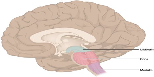
The medulla oblongata, also known just as the medulla, is part of your brainstem, which is literally the stem that extends from our brain. The medulla sits below the pons and above the spinal cord and is a major relay point for information going to and from our brain and spinal cord. In fact, its ‘middle-man’ position is actually reflected in its name, which means ‘elongated’ or ‘oblong’ (oblongata) ‘middle’ (medulla).
The bulb is an archaic term for the medulla oblongata and in modern clinical usage the word bulbar is retained for terms that relate to the medulla oblongata, particularly in reference to medical conditions. The word bulbar can refer to the nerves and tracts connected to the medulla, and also by association to those muscles innervated, such as those of the tongue, pharynx and larynx.
Structure and Functions of Medulla Oblongata
Medulla oblongata is connected by the pons to the midbrain and is continuous posteriorly with the spinal cord, with which it merges at the opening (foramen magnum) at the base of the skull. The medulla is divided into two main parts: the ventral medulla and the dorsal medulla; also known as the tegmentu. The ventral medulla contains a pair of triangular structures called pyramids, within which lie the pyramidal tracts. The pyramidal tracts are made up of the corticospinal tract and the corticobulbar tract. In their descent through the lower portion of the medulla (immediately above the junction with the spinal cord), the vast majority (80 to 90 percent) of corticospinal tracts cross, forming the point known as the decussation of the pyramids. The ventral medulla also houses another set of paired structures, the olivary bodies, which are located laterally on the pyramids.
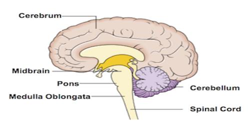
The posterior part of the medulla between the posterior median sulcus and the posterolateral sulcus contains tracts that enter it from the posterior funiculus of the spinal cord. These are the gracile fasciculus, lying medially next to the midline, and the cuneate fasciculus, lying laterally. These fasciculi end in rounded elevations known as the gracile and the cuneate tubercles. They are caused by masses of gray matter known as the gracile nucleus and the cuneate nucleus. The soma (cell bodies) in these nuclei are the second-order neurons of the posterior column-medial lemniscus pathway, and their axons, called the internal arcuate fibers or fasciculi, decussate from one side of the medulla to the other to form the medial lemniscus.
The medulla oblongata helps regulate breathing, heart and blood vessel function, digestion, sneezing, and swallowing. This part of the brain is a center for respiration and circulation. Sensory and motor neurons (nerve cells) from the forebrain and midbrain travel through the medulla. The main function of the thalamus is to process information to and from the spinal cord and the cerebellum.
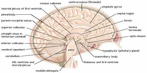
The medulla oblongata receives its blood supply from several arteries, including the anterior spinal artery, posterior inferior cerebellar artery, and the vertebral artery’s direct branches. It contains both myelinated and unmyelinated nerve fibers, also called white matter and gray matter, respectively.
Both lampreys and hagfish possess a fully developed medulla oblongata. Since these are both very similar to early agnathans, it has been suggested that the medulla evolved in these early fish, approximately 505 million years ago. The status of the medulla as part of the primordial reptilian brain is confirmed by its disproportionate size in modern reptiles such as the crocodile, alligator, and monitor lizard.
Reference:
