Inferior Vena Cava (IVC)
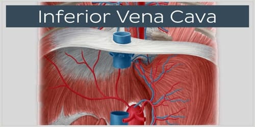
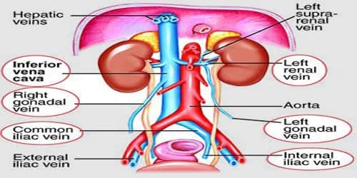
Definition: Inferior vena cava (or IVC) is a large vein that carries de-oxygenated blood from the lower body to the heart. It is also referred to as the posterior vena cava. Its walls are rigid and have valves so the blood does not flow down via gravity. It is formed by the joining of the right and the left common iliac veins, usually at the level of the fifth lumbar vertebra.
The inferior vena cava empties into the right atrium of the heart. The right atrium is located on the lower right back side of the heart. The inferior vena cava runs posterior, or behind, the abdominal cavity. This vein also runs alongside the right vertebral column of the spine.
The inferior vena cava is the lower (“inferior”) of the two venae cavae, the two large veins that carry deoxygenated blood from the body to the right atrium of the heart: the inferior vena cava carries blood from the lower half of the body whilst the superior vena cava carries blood from the upper half of the body. Together, the venae cavae (in addition to the coronary sinus, which carries blood from the muscle of the heart itself) form the venous counterparts of the aorta.
Below is a list of (most common) vertebral levels at which different veins enter the IVC.
- T8: Hepatic veins, inferior phrenic veins
- L1: Right suprarenal vein, renal veins
- L2: Left gonadal vein
- L1-L5: Lumbar vertebral veins
- L5: Right and left common iliac veins
The inferior vena cava is the result of two major leg veins coming together. These leg veins are called iliac veins. The iliac veins come together at the small of the back, at the fifth lumbar vertebra. Once the iliac veins have merged, the inferior vena cava begins to transport blood to the heart.
It is a large retroperitoneal vein that lies posterior to the abdominal cavity and runs along the right side of the vertebral column. It enters the right atrium at the lower right, the back side of the heart. The name derives from Latin: vena, “vein”, cavus, “hollow.
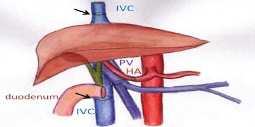
Structure and Functions of Inferior vena cava: The inferior vena cava is ultimately responsible for the transport of almost all venous blood (deoxygenated) from the abdomen and lower extremities back to the right side of the heart for oxygenation.
The IVC is formed by the confluence of the left and right common iliac veins. Numerous paired segmental lumbar veins drain into the IVC throughout its length. The right gonadal vein empties directly into the cava, whereas the left gonadal vein generally empties into the left renal vein. The azygous system has connections with the IVC or the renal veins at the level of the renal veins.
The next major veins encountered are the renal veins, followed by the hepatic veins. No valves are within the IVC. The cava enters the thoracic cavity through the tendinous portion of the diaphragm and terminates at its junction with the right atrium.
The normal diameter of the IVC is 1.5-2.5cm – this varies depending on inspiration and expiration and also with the patient’s volume status. A diameter of <1cm indicates hypovolaemia, whereas >2.5cm suggests fluid overload.
As the central venous pressure is normally very low (5-10mmHg), IVC aneurysms are exceptionally rare. The low pressure instead makes it vulnerable to obstruction, which can be due to internal occlusion by thrombosis or spreading cancer (“tumor thrombus”), or external compression by an aortic aneurysm, intra-abdominal malignancies, or a heavily pregnant uterus.
In the embryo, the inferior vena cava and right atrium are separated by the valve of the inferior vena cava, also known as the Eustachian valve. In the adult, this valve typically has totally regressed or remains as a small fold of endocardium.
Rarely, the inferior vena cava may vary in its size and position. In transposition of the great arteries, the inferior vena cava may lie on the left. It may be replaced by two vessels beneath the level of the renal veins.
The IVC does not drain blood from the gut. This has to pass through the portal vein into the liver, to allow removal of any contaminants and processing of the nutrients. The portal vein is formed by the union of the splenic vein and the superior mesenteric vein behind the neck of the pancreas. It travels into the liver as part of the portal triad in the lower free edge of the lesser omentum. Once processed, venous blood passes back into the systemic circulation via the three hepatic veins.
Health problems attributed to the IVC are most often associated with it being compressed (ruptures are rare because it has a low intraluminal pressure). Typical sources of external pressure are an enlarged aorta (abdominal aortic aneurysm), the gravid uterus (aortocaval compression syndrome) and abdominal malignancies, such as colorectal cancer, renal cell carcinoma, and ovarian cancer. Since the inferior vena cava is primarily a right-sided structure, unconscious pregnant women should be turned on to their left side (the recovery position), to relieve pressure on it and facilitate venous return. In rare cases, straining associated with defecation can lead to restricted blood flow through the IVC and result in syncope (fainting).
Information Source:
