A novel endoscope that combines photoacoustic and fluorescence imaging in a device roughly the thickness of a human hair has been demonstrated by researchers. By allowing blood dynamics to be studied at the same time as neuronal activity, the device could one day reveal fresh insights into the brain.
“Combining these imaging modalities could improve our understanding of the brain’s structure and behavior in specific conditions such as after treatment with a targeted drug,” said research team leader Emmanuel Bossy from the CNRS/ Université Grenobe Alpes Laboratoire Interdisciplinaire de Physique.
“The endoscope’s small size helps minimize damage to tissue when inserting it into the brains of small animals for imaging.”
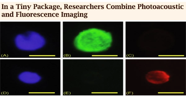
Bossy’s research team describes their new multi-modality endoscope in the Optical Society (OSA) journal Biomedical Optics Express, in collaboration with Paul C. Beard’s team from University College London, and shows that it can acquire photoacoustic and fluorescent images of red blood cells and fluorescent beads.
Two images are better than one
Using the same instrument to capture fluorescence and photoacoustic images results in automatically co-registered images with complementary information. Fluorescent signals are most useful for marking specific sections of tissue because they are formed when a fluorescent marker absorbs light and re-emits it with a different wavelength.
Light propagating into a multi-mode fiber is scrambled, making it impossible to see through the fiber. However, this type of fiber is advantageous for endoscopy because it is extremely small compared to the bundles of imaging fibers used for many medical endoscopic devices.
Emmanuel Bossy
Photoacoustic pictures, on the other hand, which capture an acoustic wave generated when light is absorbed, do not require labels and can thus be used to image blood dynamics, for example. The novel endoscope creates a focused patch of light at the imaging tip of a very thin multi-mode optical fiber using a process called optical wavefront shaping.
“Light propagating into a multi-mode fiber is scrambled, making it impossible to see through the fiber,” said Bossy. “However, this type of fiber is advantageous for endoscopy because it is extremely small compared to the bundles of imaging fibers used for many medical endoscopic devices.”
The researchers employed a spatial light modulator to send specific light patterns across the fiber and establish a focus spot at the imaging end to look through the multi-mode optical fiber. When the focus spot strikes the sample, it generates a signal that may be utilized to raster scan the spot over the sample to build up a picture point by point.
Although other researchers have employed multi-mode fibers for fluorescence endoscopy, this is the first time photoacoustic imaging has been included into an endoscopic design of this type.
Adding sound sensitivity
By integrating an additional, very thin optical fiber with a particular sound-sensitive sensor tip, the researchers were able to add photoacoustic imaging. The researchers employed a highly sensitive fiber-optic sensor built by Beard’s research team because commercially available fiber-optic acoustic sensors aren’t sensitive or tiny enough for this application.
“The focused spot of light allows us to build the image pixel by pixel while also increasing the strength of fluorescence and photoacoustic signals because it concentrates the light at the focal spot,” explained Bossy.
“This concentrated light combined with a sensitive detector made it possible to obtain images using only one laser pulse per pixel, whereas commercial fiber optic acoustic sensors would have required many laser pulses.”
The researchers created a prototype microendoscope that measured just 250 by 125 microns cubed and employed both imaging modalities to image fluorescent beads and blood cells. Multiple 1-micron fluorescent beads and individual 6-micron red blood cells were successfully detected.
The researchers are optimistic that their dual-modality gadget will operate in comparable situations because other scientists have performed fluorescence endoscopy in mouse brains. They’re now working to speed up the device’s acquisition, with the objective of capturing a few photographs each second.