Before Jeremy Schmahmann and Janet Sherman of Harvard’s Massachusetts General Hospital published a paradigm-shifting paper in the journal Brain in 1998, the majority of neuroscientists believed that the cerebellum was a motor-only brain region that coordinated muscle movements but had nothing to do with cognition or social behaviors.
For instance, my neuroscientist father, who also worked at Harvard Medical School, mentored me as a beginner tennis player in the 1970s and incorporated cerebellar functions into my tennis training.
Dad explained to me how Purkinje cells in the cerebellum store muscle memory, enabling athletes to hit home runs, kick field goals, and serve aces without consciously planning their movements.
Dad was a tennis instructor and understood that a player’s chances of performing at their best on the court increased if they developed their so-called “little brain” to automatically execute precise muscular motions.
Additionally, he was aware that improving cerebellar functions promoted superfluidity, a condition in which fluids flow without resistance, and reduced the likelihood that a tennis player would make a mistake by overanalyzing each shot.
In addition, because he was both a neuroscientist and a neurosurgeon, my father was able to operate on other people’s brains with ease under pressure because to his metacognitive understanding of the function that Purkinje cells play in muscle memory and preventing choking.
Dad also linked enhancing cerebellar functions with improving his abilities as a better brain surgeon in the operating room, much how “practice, practice, practice” improved his cerebellum’s capacity to serve aces and prevent unforced errors on the tennis court.
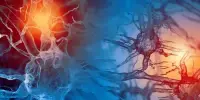
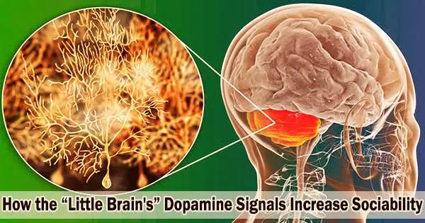
We have uncovered an unexpected causal link between Purkinje cells’ dopamine D2R expression levels right in the center of the cerebellum, the Crus I/II lobules, and the modulation of social behaviors.
Laura Cutando
My father was a 20th century brain surgeon who operated on patients with tumors, lesions, or damage to various areas of the cerebellum. He observed that some patients’ cerebellar anomalies did not appear to impede motor functions, but did influence them in other ways during the 1970s, ’80s, and ’90s. These observations were confusing since no one believed at the time that the cerebellum was involved in non-motor processes.
The Cerebellum’s Role in Social Cognition Remains a Mystery
When Dad served as a medical consultant for my book The Athlete’s Way in 2005, he encouraged me to put the cerebellum and Purkinje cells in the spotlight because of their well-established role in motor processes and athletic mastery of complex motor sequences.
He also recommended that any brain maps of cerebro-cerebellar circuitry included in the book should represent that the cerebellum was more than just a motor-function-only brain center based on his first-hand observations of cerebellar abnormalities affecting social and emotional processes and accumulating evidence-based research.
However, nobody was fully aware in the aughts that diverse cerebellar microzones and regions were involved in both motor and non-motor processes. Dad’s way of summing up all of the cerebellar unknowns circa 2005 was to say, “We don’t know exactly what the cerebellum is doing, but whatever it’s doing, it’s doing a lot of it.”
It wasn’t until about 2015 that neuroscientists began to zero in on how different regions and microzones of the cerebellum play a role in social cognition.
“Research on the relationship between the cerebellum and social cognition is very young, and apart from occasional early contributions, began to emerge over the past five years,” Frank Van Overwalle and Mario Manto wrote in 2020. “Prior reports on the social role of the cerebellum were often limited to side aspects of affective processing and anecdotally described cerebellar patients having affective deficits.”
Dopamine D2 Receptors in the Cerebellum May Regulate Social Behavior
Neuroscientists have been attempting to determine how the cerebellum affects social behavior in both humans and animals for the past ten years.
A new animal study (Cutando et al., 2022) by an international team of researchers sheds light on how dopamine D2 receptors (D2Rs) regulate social behavior in mice. These findings were published on June 16 in the peer-reviewed journal Nature Neuroscience.
“We have uncovered an unexpected causal link between Purkinje cells’ dopamine D2R expression levels right in the center of the cerebellum, the Crus I/II lobules, and the modulation of social behaviors,” first author Laura Cutando of Universitat Autònoma de Barcelona (UAB) said in a June 2022 news release.
“Reducing the expression of this specific dopamine receptor impaired the sociability of mice as well as their preference for social novelty, while their coordination and motor functions remained unaffected,” she wrote.
Cutando et al. used genetic tools and different mouse models to investigate how altering D2R levels in cerebellar Purkinje cells could modify social behaviors without affecting motor functions.
By selectively modifying dopamine D2 signaling in a mouse’s Purkinje cells, the researchers could analyze how under and over-expression of D2Rs affected motor and non-motor cerebellar functions. As mentioned, reducing dopamine D2R levels of expression made mice less social, whereas increasing D2R signaling increased sociability.
These results, say the researchers, could help us learn more about how dopamine signaling in the cerebellum affects psychiatric disorders like schizophrenia, attention-deficit/hyperactivity disorder (ADHD), and generalized anxiety disorder (GAD). Of course, human studies are needed to identify how cerebellar dopamine D2 receptors regulate social behaviors in people.