There are a few different technologies that could potentially be used to create “stickers” that can see inside the body, including infrared imaging, ultrasound, and magnetic resonance imaging (MRI). These technologies have been used in the past to create portable and non-invasive medical devices that can be used to image the inside of the body.
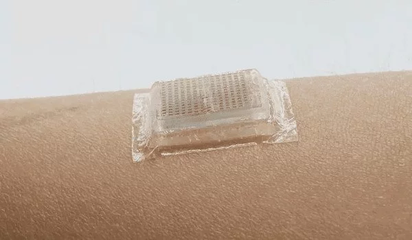
Engineers created an adhesive patch that generates ultrasound images of the human body. The stamp-sized device adheres to the skin and can provide 48 hours of continuous ultrasound imaging of internal organs. Ultrasound imaging provides clinicians with live images of a patient’s internal organs and is a safe and noninvasive window into the body’s workings. Trained technicians use ultrasound wands and probes to direct sound waves into the body in order to capture these images. These reflected waves produce high-resolution images of a patient’s heart, lungs, and other deep organs.
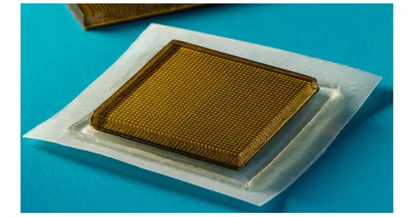
Currently, ultrasound imaging necessitates the use of large, specialized equipment that is only available in hospitals and doctor’s offices. However, a new design developed by MIT engineers may make the technology as wearable and accessible as buying Band-Aids at the pharmacy. The engineers present the design for a new ultrasound sticker in Science, a stamp-sized device that sticks to the skin and can provide continuous ultrasound imaging of internal organs for 48 hours.
Wearable ultrasound imaging tool would have huge potential in the future of clinical diagnosis. However, the resolution and imaging duration of existing ultrasound patches is relatively low, and they cannot image deep organs.
Chonghe Wang
The stickers were applied to volunteers, and the devices produced live, high-resolution images of major blood vessels and deeper organs such as the heart, lungs, and stomach. The stickers stayed put and recorded changes in underlying organs as volunteers did various activities like sitting, standing, jogging, and biking.
According to the current design, the stickers must be connected to instruments that convert the reflected sound waves into images. The researchers point out that the stickers, even in their current form, could have immediate applications: For example, similar to heart-monitoring EKG stickers, the devices could be applied to patients in the hospital and continuously image internal organs without requiring a technician to hold a probe in place for long periods of time.
If the devices can be made to operate wirelessly – a goal the team is currently working toward – the ultrasound stickers could be made into wearable imaging products that patients could take home from a doctor’s office or even buy at a pharmacy.
“We envision a few patches adhered to different locations on the body, and the patches would communicate with your cellphone, where AI algorithms would analyze the images on demand,” says the study’s senior author, Xuanhe Zhao, professor of mechanical engineering and civil and environmental engineering at MIT. “We believe we’ve opened a new era of wearable imaging: With a few patches on your body, you could see your internal organs.”
The study also includes lead authors Chonghe Wang and Xiaoyu Chen, and co-authors Liu Wang, Mitsutoshi Makihata, and Tao Zhao at MIT, along with Hsiao-Chuan Liu of the Mayo Clinic in Rochester, Minnesota.
A sticky issue
A technician first applies a liquid gel to a patient’s skin, which acts as a conduit for ultrasound waves. After that, a probe, or transducer, is pressed against the gel, sending sound waves into the body that reverberate off internal structures and back to the probe, where the echoed signals are translated into visual images.
Some hospitals offer probes attached to robotic arms that can hold a transducer in place without tiring, but the liquid ultrasound gel flows away and dries out over time, interfering with long-term imaging.
Researchers have been investigating designs for stretchable ultrasound probes that would provide portable, low-profile imaging of internal organs in recent years. These designs provided a flexible array of tiny ultrasound transducers, with the idea that such a device would stretch and conform to the body of a patient. However, due to their stretch, these experimental designs produced low-resolution images: Transducers shift relative to each other as they move with the body, distorting the resulting image.
“Wearable ultrasound imaging tool would have huge potential in the future of clinical diagnosis. However, the resolution and imaging duration of existing ultrasound patches is relatively low, and they cannot image deep organs,” says Chonghe Wang, who is an MIT graduate student.
An inside look
By combining a stretchy adhesive layer with a rigid array of transducers, the MIT team’s new ultrasound sticker produces higher-resolution images for a longer period of time. “This combination allows the device to conform to the skin while maintaining transducer relative location to generate clearer and more precise images.” Wang claims.
The adhesive layer of the device is composed of two thin layers of elastomer that encapsulate a middle layer of solid hydrogel, a mostly water-based material that easily transmits sound waves. The MIT team’s hydrogel is elastic and stretchy, unlike traditional ultrasound gels.
“The elastomer prevents dehydration of hydrogel,” says Chen, an MIT postdoc. “Only when the hydrogel is highly hydrated can acoustic waves penetrate effectively and give high-resolution imaging of internal organs.”
The bottom elastomer layer is designed to stick to skin, while the top layer adheres to a rigid array of transducers that the team also designed and fabricated. The entire ultrasound sticker measures about 2 square centimeters across, and 3 millimeters thick – about the area of a postage stamp.
The researchers ran the ultrasound sticker through a battery of tests with healthy volunteers, who wore the stickers on various parts of their bodies, including the neck, chest, abdomen, and arms. The stickers stayed attached to their skin, and produced clear images of underlying structures for up to 48 hours. During this time, volunteers performed a variety of activities in the lab, from sitting and standing, to jogging, biking, and lifting weights.
The team was able to observe the changing diameter of major blood vessels when seated versus standing using the images from the stickers. The stickers also captured information about deeper organs, such as how the heart changes shape during exercise. The researchers could also see the stomach expand and then contract as volunteers drank and then passed juice out of their systems. The team was also able to detect bright patterns in underlying muscles as some volunteers lifted weights, indicating temporary microdamage.
“With imaging, we might be able to capture the moment in a workout before overuse, and stop before muscles become sore,” says Chen. “We do not know when that moment might be yet, but now we can provide imaging data that experts can interpret.”
The team is working on making the stickers wireless. They are also working on artificial intelligence-based software algorithms to better interpret and diagnose the images of the stickers. Then, Zhao envisions ultrasound stickers being packaged and purchased by patients and consumers and being used to monitor not only various internal organs, but also tumor progression and fetal development in the womb.
“We envision a box of stickers, each designed to depict a different part of the body,” Zhao says. “We believe this represents a significant advancement in wearable devices and medical imaging.”