Peripheral vascular disease (PVD) refers to a group of conditions that affect the blood vessels outside the heart and brain, typically those in the legs and feet. These conditions are caused by a buildup of plaque in the arteries, which can lead to reduced blood flow and oxygen delivery to the affected area.
With a plethora of peripheral blood arteries connecting the heart to the limbs, our body resembles a complex network. A wound may not heal or may result in necrosis if blood is unable to flow through these peripheral blood veins.
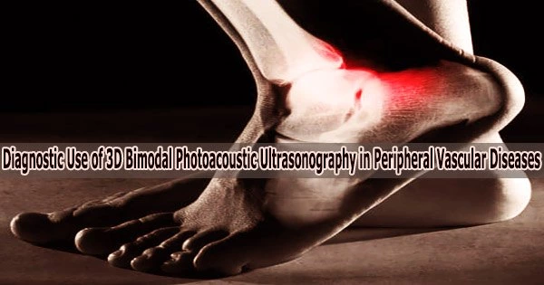
Peripheral vascular illnesses need special care since they typically affect the foot, which is essential to one’s health and ability to function. Recently, a Korean research team has developed a 3D foot imaging technique that vividly captures peripheral blood vessels, even thinner than 1 mm.
A POSTECH research team led by Professor Chulhong Kim (Belonging to the Departments of Convergence IT Engineering, Electrical Engineering, and Mechanical Engineering) has developed an imaging technique that combines the photoacoustic and ultrasound images. The findings from the study were recently published in the international journal Radiology.
The traditional method for diagnosing peripheral vascular illnesses used the ankle-brachial index test to monitor the blood pressure in the wrist, ankle, or toe and determine the ratio. Doppler ultrasonography, which monitors blood flow, angiography utilizing computed tomography (CT), and magnetic resonance imaging (MRI) were the most popular imaging modalities.
These techniques have limits in their ability to clearly capture the thin and many peripheral blood capillaries, even though they can detect anomalies in main arteries. Because a contrast agent needs to be injected into the patient, they may also cause discomfort or unwanted effects.
The researchers combined photoacoustic and ultrasound pictures to visualize 3D images of blood arteries thinner than 1 mm without the need of a contrast agent in order to get around these problems.
Blood vessels in the body can be observed via the photoacoustic effect, in which sound waves are created after light absorption in a substance, without the use of a contrast agent. When the ultrasound picture is added, the structural image of the blood vessels, the image of the skin, and the image of the bone structures may all be seen at once.
The new modality can provide the information to aid in diagnosing and treating peripheral vascular diseases since it provides functional diagnostic values for the blood supply to tissues, such as total hemoglobin concentration or blood oxygen saturation, using a wavelength-convertible laser.
The researchers also created a contour scan technique, which uses the imaging probe to detect the foot’s contours and travel along them, to increase the consistency and reliability of picture data.
The researchers experimented on participants’ healthy feet to demonstrate its utility as a diagnostic tool. The ability to simultaneously display the foot’s blood arteries, skin, and bone structures was confirmed by taking photoacoustic and ultrasound images of the entire foot in action.
Also, the researchers artificially restricted the peripheral blood flow in the healthy volunteers using a pressure cuff, and they noted substantial alterations in hemoglobin concentration and vessel density before and after the occlusion.
The researchers have confirmed the novel imaging technology’s potential use for identifying peripheral vascular disorders in the future based on the findings of this investigation.
This study was conducted with the support from the Ministry of Science and ICT, Ministry of Education, and the National Research Foundation of Korea.