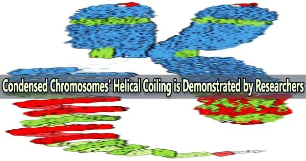
Condensed chromosomes refer to the highly compacted and tightly coiled form of DNA that is visible during cell division. Chromosomes undergo condensation during mitosis or meiosis, which allows them to be efficiently separated and distributed to daughter cells.
Early cytological research revealed that the metaphase chromosomes’ chromatids form a spiral structure called a chromonema. Chromosome conformation capture sequencing has recently provided evidence in favor of this hypothesis. Yet, there was no direct observation of the coiled chromonema to support the helical concept.
Now, for the first time an international research team led by the IPK Leibniz Insititute and the Institute of Experimental Botany of the Czech Academy of Science provides the direct proof of the helical coiling of condensed chromatids via super-resolution microscopy of specifically labelled chromonema regions. Today, the results have been published in the journal Nucleic Acids Research.
In textbooks and other media, the famous X-shaped arrangement of metaphase chromosomes is commonly depicted. The illustrations provide an interesting explanation of how chromosomes retain the majority of genetic information and pass it on to the following generation.
“These presentations suggest that the chromosome ultrastructure is well-understood. However, this is not the case,” says Dr. Veit Schubert from IPK’s research group “Chromosome structure and function.”
To study the higher-order structure of mitotic chromosomes, the large chromosomes of barley (Hordeum vulgare) were used as a model. A single helical turn covers 20-38 Mb, creating a ~400 nm thick fiber, which we identify as the chromonema.
Dr. Amanda Camara
Based on data gathered using a variety of molecular and microscopy methods, several models have been put forth to characterize the higher-order structure of metaphase chromosomes. Both helical and non-helical versions of these models are recognized.
Whereas non-helical theories propose that chromatin is folded within the chromatids without creating a spiral, helical models believe that the chromatin in each sister chromatid at metaphase is structured as a coil.
The researchers revived the term ‘chromonema’ which was used for the first time at the beginning of the 20th century. Now, the IPK and IEB researchers provided a detailed description of its ultrastructure.
An independent proof for the coiling of the chromonema was provided by a variety of experimental methods, including chromosome conformation capture sequencing (Hi-C) of isolated mitotic chromosomes, polymer modeling, microscopic observations of sister chromatid exchanges, and oligo-FISH labelled regions at the super-resolution level.
“Our multidisciplinary approach demonstrates that the coiled chromatid organization and its organizational unit, the chromonema, can be confirmed independently by different methods.” says Dr. Veit Schubert.
Condensed chromosomes are essential for proper cell division and the accurate transmission of genetic information from parent to daughter cells. Without proper condensation, chromosomes can become tangled and broken, leading to chromosomal abnormalities and genetic disorders.
“To study the higher-order structure of mitotic chromosomes, the large chromosomes of barley (Hordeum vulgare) were used as a model. A single helical turn covers 20-38 Mb, creating a ~400 nm thick fiber, which we identify as the chromonema,” says Dr. Amanda Camara, one of the frist authors of the study.
The concept suggests a universal mechanism for condensed mitotic chromosome production that can be used by all eukaryotes with a wide range of genome sizes.
“We expect that following our study, chromonema coiling will be confirmed in a larger number of plant and animal species containing large chromosomes. The identification of the principle of chromosome condensation in this work is the stepping stone to understanding chromatin dynamics during the course of the cell cycle,” says Dr. Amanda Camara.