Auditory Cortex
Definition
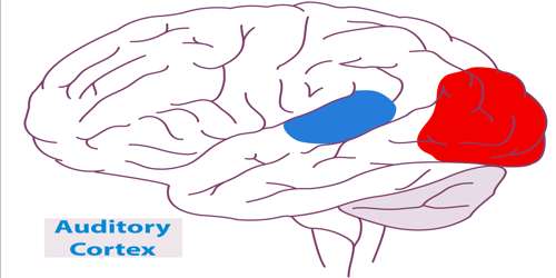
Auditory cortex is the section of the brain that processes information received through hearing. It is also called auditory area. It is located bilaterally, roughly at the upper sides of the temporal lobes – in humans on the superior temporal plane, within the lateral fissure and comprising parts of Heschl’s gyrus and the superior temporal gyrus, including planum polare and planum temporale (roughly Brodmann areas 41, 42, and partially 22). Unilateral destruction results in slight hearing loss, whereas bilateral destruction results in cortical deafness.
Auditory cortex, narrower in humans than in other mammals, develops from front to back within the Sylvian fissure at the point where it joins Heschl’s gyrus.
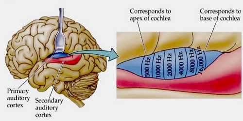
- Primary auditory cortex (AI) is situated in the posterior third of the superior temporal gyrus (also known as Brodmann area 41), next to Wernicke’s area (W). AI is the central region of the auditory cortex and receives direct projections from the ascending auditory pathway, particularly the ventral region of the medial geniculate body (MGB) in the thalamus.
- Secondary auditory cortex (AII) is located more rostrally in the temporal lobe and contains Brodmann area 42.
Structure and Functions of Auditory Cortex
The presence of six cell layers in the auditory cortex is common to all mammals, but species differences take the form of the commonality of each cells within each layer. In humans, pyramidal cells (including all types) correspond to 85% of AI. The remaining 15% are multipolar or stellate cells. Inverted stellate cells also exist (Martinotti cells) as well as cells with candelabra-shaped dendritic configurations.

Like many areas in the neocortex, the functional properties of the adult primary auditory cortex (A1) are highly dependent on the sounds encountered early in life. This has been best studied using animal models, especially cats and rats. In the rat, exposure to a single frequency during postnatal day (P) 11 to 13 can cause a 2-fold expansion in the representation of that frequency in A1. Importantly, the change is persistent, in that it lasts throughout the animal’s life, and specific, in that the same exposure outside of that period causes no lasting change in the tonotopy of A1.
Neurons in AI and AII are functionally organized into columns, first described by Lorent de Nó. Cortical columns receive input from both MGBs and are therefore bilateral, working on the principal of summation/suppression. Summation corresponds to similar afferentation from both ears, with a contralateral dominance. Suppression is ipsilaterally dominant. Each neuron of the MGB that projects to the auditory cortex (C) generates a fiber (f-1) that branches horizontally for a few millimeters and contacts numerous pyramidal cells (B) and puncta (C). This system allows the amplification of the auditory signal and improved analysis of its activity.
The auditory cortex plays an important yet ambiguous role in hearing. When the auditory information passes into the cortex, the specifics of what exactly takes place are unclear. There is a large degree of individual variation in the auditory cortex, as noted by biologist James Beament, who wrote, “The cortex is so complex that the most we may ever hope for is to understand it in principle, since the evidence we already have suggests that no two cortices work in precisely the same way.”
While other levels of the auditory pathways are very similar within species, the human neocortex is characterized by the predominence of pyramidal cells (85% of cortical neurons), and some very specific types of cells such as inverted pyramidal cells and candelabrum neurons. Another specificity is the massive interconnection of cortical neurons, which accounts for 80% of excitatory synapses in the neocortex.
Reference: