Tumor formation begins with minute alterations within the body’s cells; ion diffusion at the smallest scales is critical in battery function. Until now, the resolution of traditional imaging technologies has not been high enough to accurately capture these processes. A research team led by the Technical University of Munich (TUM) has created diamond quantum sensors that can be used to improve magnetic imaging resolution.
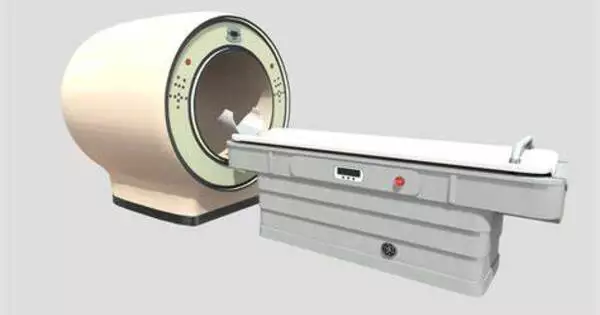
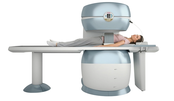
Nuclear magnetic resonance (NMR) is a valuable imaging technique in research that may be used to see tissue and structures without causing damage to them. Magnetic Resonance Imaging (MRI) is a medical technique in which the patient is moved into a bore of a huge magnet on a table. The MRI machine generates a very strong magnetic field that interacts with the small magnetic fields of the body’s hydrogen nuclei. Because hydrogen atoms are dispersed differently in different types of tissues, it is feasible to distinguish organs, joints, muscles, and blood vessels.
NMR technologies can also be utilized to visualize water and other element diffusion. Observing the behavior of carbon or lithium, for example, is frequently used in research to investigate the architecture of enzymes or processes in batteries. “Existing NMR methods provide good results, for example, when it comes to recognizing abnormal processes in cell colonies,” says TUM Professor for Quantum Sensing Dominik Bucher. “But we need new approaches if we want to explain what happens in the microstructures within the single cells.”
The ability of NMR and MRI techniques to directly detect the mobility of atoms and molecules makes them absolutely unique when compared to other imaging methods. We now have found a way, how their spatial resolution, which is currently often deemed insufficient, can be significantly improved in future.
Prof. Maxim Zaitsev
Sensors made of diamond
For this objective, the study team created a quantum sensor constructed of synthetic diamond. “We enrich the diamond layer, which we provide for the new NMR method, with special nitrogen and carbon atoms already during growth,” Dr. Peter Knittel of the Fraunhofer Institute for Applied Solid State Physics (IAF) says.
Electron irradiation after growth separates individual carbon atoms from the diamond’s flawless crystal lattice. The resultant defects arrange themselves next to the nitrogen atoms, forming a nitrogen-vacancy center. These vacancies have unique quantum mechanical features that are required for sensing. “Our processing of the material optimizes the duration of the quantum states, which allows the sensors to measure for longer,” Knittel said.
Quantum sensors pass the first test
A diamond-based tiny magnetic resonance imager (MRI) is an intriguing concept with potential benefits, but it faces significant technological obstacles. An MRI machine is normally enormous and expensive, however using diamond in a small MRI could provide unique benefits due to the material’s characteristics.
Magnetic fields interact with the quantum state of nitrogen-vacancy centers. “The MRI signal from the sample is then converted into an optical signal which we can detect with a high degree of spatial resolution,” Bucher said.
TUM scientists put a microchip with small water-filled channels on the diamond quantum sensor to test it. “This allows us to simulate microstructures of a cell,” Bucher says. The researchers were able to successfully evaluate the migration of water molecules within the microstructure.
In the following stage, the researchers hope to improve the technology so that it may be used to investigate microstructures in single living cells, tissue sections, or the ion mobility of thin-film materials for battery applications. “The ability of NMR and MRI techniques to directly detect the mobility of atoms and molecules makes them absolutely unique when compared to other imaging methods,” explains Prof. Maxim Zaitsev of the University of Freiburg. “We now have found a way, how their spatial resolution, which is currently often deemed insufficient, can be significantly improved in future.”