We know a lot about Tyrannosaurus rex jaws, from the bite force of a baby dinosaur to how adults crunched on their prey like an alligator. Now, medical researchers have used a CT (computed tomography) scan to reveal that these ancient carnivores had bone disease. A clinical CT scanner was used to examine the 68-million-year-old lower jawbone of a T-rex skeleton in a new study led by radiologist Charlie Hamm from Charité University Hospital in Berlin.
According to a study presented today at the annual meeting of the Radiological Society of North America, researchers in Germany identified the bone disease in the fossilized jaw of a Tyrannosaurus rex using a CT-based, nondestructive imaging approach (RSNA). According to the researchers, the imaging method could have significant applications in paleontology as an alternative to fossil assessment methods that involve the destruction of samples.
The T. rex, a well-known subject in today’s popular culture, was a massive, carnivorous dinosaur that roamed what is now the western United States millions of years ago. One of the most complete T. rex skeletons ever discovered was discovered in Carter County, Montana, in 2010 by a commercial paleontologist. The fossilized skeleton dates back to the Late Cretaceous period, approximately 68 million years. It was sold to an investment banker, who dubbed it “Tristan Otto” before loaning it to Germany’s Museum für Naturkunde Berlin. It is one of Europe’s only two original T. rex skeletons.
We hypothesized that DECT could potentially allow for quantitative noninvasive element-based material decomposition, assisting paleontologists in characterizing unique fossils.
Charlie Hamm
Charlie Hamm, M.D., a radiologist at Berlin’s Charité University Hospital, and his colleagues recently had the opportunity to examine a portion of Tristan Otto’s lower left jaw. While previous fossil studies have mostly relied on invasive sampling and analysis, Dr. Hamm and colleagues used a noninvasive approach with a clinical CT scanner and a technique known as dual-energy computed tomography (DECT). DECT uses X-rays at two different energy levels to provide information about tissue composition and disease processes that single-energy CT cannot.
“We hypothesized that DECT could potentially allow for quantitative noninvasive element-based material decomposition, assisting paleontologists in characterizing unique fossils,” Dr. Hamm explained.
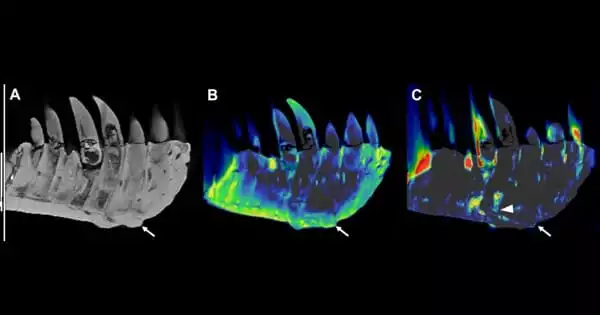
The CT technique allowed the researchers to overcome the challenges of scanning a large portion of Tristan Otto’s lower jaw, known as the left dentary. Because of the piece’s high density, CT imaging quality is known to suffer from artifacts, or misrepresentations of tissue structures, when looking at very dense objects.
“We needed to adjust the CT scanner’s tube current and voltage in order to minimize artifacts and improve image quality,” Dr. Hamm said.
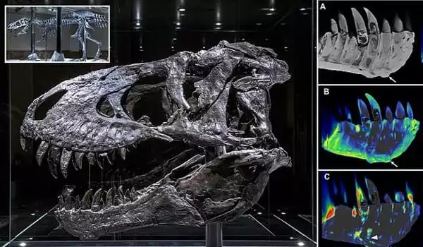
On visual inspection and CT imaging, the left dentary showed thickening and a mass on its surface that extended to the root of one of the teeth. DECT detected a significant accumulation of the element fluorine in the mass, which was associated with areas of decreased bone density. The mass and fluorine accumulation supported the diagnosis of tumefactive osteomyelitis, a bone infection.
Co-author of the study, vertebrate palaeontologist Oliver Hampe of the Natural History Museum in Berlin, agrees.
“The DECT approach has promise in other paleontological applications, such as determining age and distinguishing actual bone from replicas,” he says. “The experimental design, which includes the use of a clinical CT scanner, will enable a wide range of applications.” The findings were presented at the Radiological Society of North America’s annual meeting this week.
“While this is a proof-of-concept study, noninvasive DECT imaging that provides structural and molecular information on unique fossil objects has the potential to address an unmet need in paleontology by avoiding defragmentation or destruction,” said Dr. Hamm.
“The DECT approach has promise in other paleontological applications, such as determining age and distinguishing actual bone from replicas,” said Oliver Hampe, Ph.D., senior scientist and vertebrate paleontologist at the Museum für Naturkunde Berlin. “The experimental design, which includes the use of a clinical CT scanner, will enable a wide range of applications.”
Dr. Hamm and his colleagues also worked with paleontologists from Chicago’s Field Museum and colleagues from the Richard and Loan Hill Department of Biomedical Engineering at the University of Illinois at Chicago to conduct a CT scan of the museum’s world-famous T. rex “Sue.”
“With each project, our collaborative network expanded and evolved into a truly multidisciplinary group of experts in geology, mineralogy, paleontology, and radiology, emphasizing the potential and relevance of the results to various scientific fields,” Dr. Hamm said.