Biology depends on the process of cell division, or the development of daughter cells from a mother cell. Every cell receives the same DNA and protein components that made up the cell it was descended from. However, it is still unclear exactly how these molecular building components organize themselves into new cells.
In order to study cell division, it is necessary to simultaneously observe macromolecules with sizes ranging from nanometers, such as proteins and DNA, up to millimeters, populations of cells, and across times of seconds to weeks.
Previous microscopes could only capture minute objects for little periods of time, usually just tens of seconds. There hasn’t been a method that can simultaneously investigate a variety of size and time scales.
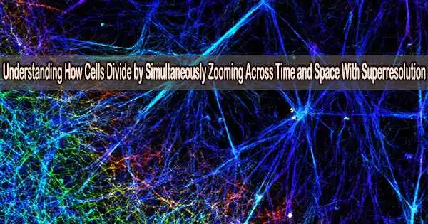
At the University of Michigan’s Bioplasmonics Group, my group and I created a brand-new sort of superresolution imaging that uncovers hitherto unobserved characteristics of cell division.
Advancing superresolution imaging
It wasn’t possible to view cells at the molecular level until recently with the 2014 Nobel Prize-winning development of superresolution.
Because light stretches out as it goes through space, conventional light microscopes distort relatively small things that are clustered together in a sample. Fluorescent probes attached to the sample could be turned on and off with superresolution, just like the stars on a starry night.
A superresolution image, created by gathering and merging numerous photographs from these probes, can reveal extremely minute objects. Superresolution revealed structures as small as 10 nanometers, or around the size of a protein molecule, opening up a whole new realm in biology.
However, the fluorescent probes that this technique relies on can quickly wear out. This restricts its application to researching lengthy processes like cell division.
My research team and I have a developed a solution we call PINE nanoscopy. The fluorescent probes we use scatter light rather than absorbing it, which prevents them from degrading under repeated light exposure.
We developed filters composed of thin layers of polymers and liquid crystals that can detect scattered light and turn on and off the probes in order to resolve very small objects that are close together. This allowed us to see nanometer-scale details of cells that would otherwise be blurred by traditional microscopes.
Remarkably, we found that these nanometer-scale details could be viewed for very long periods over 250 hours. These details would typically be lost over time with traditional superresolution methods.
Shedding new light on cell division
The organization of the molecular building blocks during cell division was then studied using our methodology.
We concentrated on a protein called actin, which serves a variety of purposes including maintaining cell structure. Actin is shaped like branching filaments, each about 7 nanometers (millionths of a millimeter) in diameter, that link together to span thousands of nanometers. Using PINE nanoscopy, we attached scattering probes to actin to visually follow human cells as they divided.
We made three observations on how actin building blocks organize during cell division. These molecular building pieces first grow to strengthen their links to one another. To enhance their points of interaction, they also become closer to their neighbors. Third, when the actin molecules are more connected to one another and when they are less attached to one another, the ensuing networks have a tendency to constrict and expand, respectively.
We were able to learn new details regarding the process of cell division as a result of these observations. We discovered that during cell division, interactions between the constituents of actin coordinate with the contraction and expansion of the entire cell. In other words, the behavior of the actin molecules is connected to the behavior of the cell: The cell contracts when the actin expands, and it expands when the actin contracts.
Uncovering disease with superresolution
Our approach will be used to investigate how different molecular building blocks arrange themselves into tissues and organs. Similar to how cells are arranged, tissues and organs follow a hierarchy that may be studied on a scale from small to enormous.
The development of new replacement tissues and organs, such as skin transplants, may benefit from examining the dynamic and intricate process of how protein building blocks interact with one another to produce larger structures.
Our imaging method will also be used to investigate how disorderly protein building components contribute to disease. Proteins form the building blocks of cells, which then form tissues, which form organs.
This organization can be disrupted by even the smallest modification in the building components, and the results may result in disorders like cancer. Our method might make it easier for researchers to see and consequently comprehend how molecular flaws in tissues and organs may result in disease.