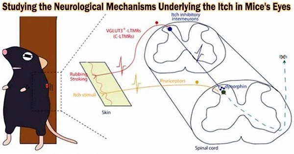
The unpleasant effects of itchy skin are well known to those who have dry skin, dermatitis, or insect bites. The physiological mechanisms that cause itching on the skin are well recognized, but the signals that cause itching in the eyes are unclear.
A research group led by Associate Professor Sakamoto Hirotaka from Okayama University (Ushimado Marine Institute; UMI) and Associate Professor Takanami Keiko from Nara Women’s University has now investigated the neural mechanisms behind itchy eyes in rodents. Additionally, the group offers information on the limb strategies used by rats to deal with this eye issue.
Their study is published in the journal Proceedings of the Royal Society B: Biological Sciences.
Itching is brought on by the chemical histamine, which is released in allergic and inflammatory diseases. A biochemical mechanism called the spinal Gastrin-releasing Peptide-Gastrin-releasing Peptide receptor (GRP-GRPR pathway) transports skin-related itch signals to neurons.
The trigeminal ganglion (TG) nerves, which carry signals from the eyes to the brain, were previously shown by the research team to contain the GRP-GRPR pathway. Thus, this system was the main target of their study.
When histamine drops were used to stimulate the rats’ eyes, the researchers initially looked at the animals’ scratching behavior. They discovered that when histamine was injected into either the left or right eye, the rats utilized the hindfoot on the same side as the problematic eye to scratch and calm it.
However, the rats preferentially used the right hindfoot for rubbing when histamine was given to cause itching in both eyes at once. When it came to scratching their eyes, the rats appeared to favor their right side.
Next, the focus moved on to biochemical pathways and the GRP-GRPR system. It was discovered that when histamine was administered to either eye, the GRPR-containing neurons on that side of the brainstem became more active. Therefore, it was plausible that the GRPR-containing neurons contributed to the transmission of itch signals from the eyes.
The scientists utilized a toxin to damage these GRPR-containing neurons in the right side of the rat brainstems in order to further corroborate this. The incidence and duration of scratching did indeed decrease after these rats received histamine drops in the eyes. The itch signals from their eyes were reduced since there weren’t any GRPR neurons that were activated.
“These findings could open up a new field of research on the mechanisms of the laterality in vertebrates and also offer new potential therapeutic approaches,” suggest the researchers.
The eye scratching test here could be used to assess the footedness of individual rodents in subsequent studies, in addition to exposing the right-sided preference animals have when using their hindlimbs.
Additionally, the role of the GRP-GRPR system in TG nerves was disclosed, and this knowledge may form the basis for developing treatments for eye disorders that induce itching.
About the gastrin-releasing peptide gastrin-releasing peptide receptor (GRP-GRPR) pathway
The eye scratching test here could be used to assess the footedness of individual rodents in subsequent studies, in addition to exposing the right-sided preference animals have when using their hindlimbs.
Additionally, the role of the GRP-GRPR system in TG nerves was disclosed, and this knowledge may form the basis for developing treatments for eye disorders that induce itching. When the two connect, biochemical signals are triggered in the brain.
Trigeminal ganglion (TG) neurons from the eyes carry GRP molecules into the bottom part of the brain. A region known as the spinal trigeminal nucleus caudalis (Sp5C) in this part of the brain is found to be rich in incoming GRP-containing nerve endings.
Moreover, the Sp5C is abundant in GRPR. To investigate the function of this pathway in eye itching, the researchers in this work used a toxin to specifically target GRPR neurons in the Sp5C.