Researchers from the University of Alabama at Birmingham have used cryo-electron microscopy to reveal the structure of a bacterial virus in unprecedented detail. This is the first known structure of a virus that can infect Staphylococcus epidermidis, and high-resolution structural information is crucial for understanding viral biology and the possibility of using the virus as a treatment for bacterial diseases.
Bacteriophages or “phages” is the terms used for viruses that infect bacteria. In conjunction with Asma Hatoum-Aslan, Ph.D., at the University of Illinois Urbana-Champaign, the UAB team led by Terje Dokland, Ph.D., has described atomic models for all or a portion of 11 different structural proteins in the phage Andhra. Science Advances published the study.
Andhra belongs to the picovirus family. The only hosts it accepts are S. epidermidis. Although largely innocuous, this skin bacteria is a major contributor to infections of indwelling medical equipment.
“Picoviruses are rarely found in phage collections and remain understudied and underused for therapeutic applications,” said Hatoum-Aslan, a phage biologist at the University of Illinois.
The advent of antibiotic resistance in Staphylococcus aureus, a similar pathogen, and S. epidermidis has rekindled interest among researchers in the possibility of utilizing bacteriophages to treat bacterial infections.
After attaching to the bacterial cell wall, breaking through it enzymatically, piercing the cell membrane, and injecting viral DNA into the cell, picoviruses inevitably cause the death of the cells they infect. They also possess other characteristics, such as a tiny genome and the inability to transfer bacterial genes between bacteria, that make them appealing candidates for therapeutic application.
It will be feasible to create customised phages using genetic engineering if one is familiar with Andhra protein structure and understands how those structures permit the virus to infect a bacteria.
“The structural basis for host specificity between phages that infect S. aureus and S. epidermidis is still poorly understood,” said Dokland, a professor of microbiology at UAB and director of the UAB Cryo-Electron Microscopy Core. “With the present study, we have gained a better understanding of the structures and functions of the Andhra gene products and the determinants of host specificity, paving the way for a more rational design of custom phages for therapeutic applications. Our findings elucidate critical features for virion assembly, host recognition and penetration.”
Because the surface teichoic acid polymers of various bacterial strains vary, staphylococcal phages normally only infect a small subset of bacteria.
With the present study, we have gained a better understanding of the structures and functions of the Andhra gene products and the determinants of host specificity, paving the way for a more rational design of custom phages for therapeutic applications. Our findings elucidate critical features for virion assembly, host recognition and penetration.
Professor Terje Dokland
“This narrow host range is a double-edged sword: On one hand, it allows the phages to target only the specific pathogen causing the disease; on the other hand, it means that the phage may need to be tailored to the patient in each specific case,” Dokland said.
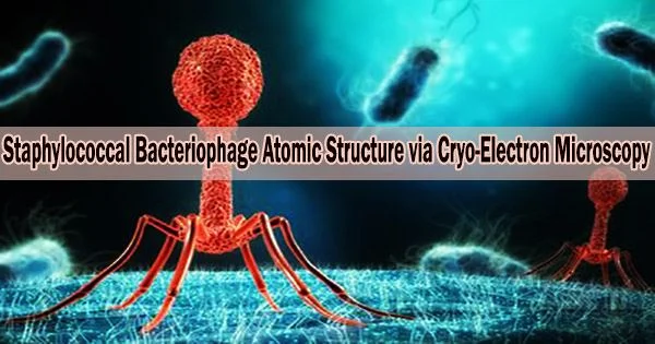
Andhra is an icosahedral capsid with 20 faces that is often roundish in shape and houses the viral genome. A brief tail is joined to the capsid. For the most part, the tail is in charge of attaching to S. epidermidis and catalyzing the breakdown of the cell wall. Through the tail, the bacterium receives the viral DNA.
The gateway from the capsid to the tail as well as the stem, appendages, knob, and tail tip are all considered to be tail segments. Each virus particle is made up of numerous copies of the 11 distinct proteins that build it up.
For instance, the nine proteins that make up a virion have copy counts ranging from two to 72, whereas the capsid is composed of 235 copies of each of its two proteins. The 12th protein, whose structure was predicted using the protein structure prediction program AlphaFold, is one of the 645 protein fragments that make up the virion in total.
The atomic models described by Dokland, Hatoum-Aslan, and co-first authors N’Toia C. Hawkins, Ph.D., and James L. Kizziah, Ph.D., UAB Department of Microbiology, show the structures for each protein as described in molecular language like alpha-helix, beta-helix, beta-strand, beta-barrel or beta-prism.
The researchers have explained how each protein interacts with neighboring proteins of different types as well as how each protein binds to other copies of that same protein type, such as to form the hexameric and pentameric faces of the capsid.
With significantly higher resolution than a light microscope, electron microscopes illuminate an item using a beam of accelerated electrons. With the addition of super-cold temperatures, cryo-electron microscopy is especially beneficial for resolving the atomic structure of bigger proteins, membrane proteins, or materials containing lipids, such as membrane-bound receptors, as well as complexes of several biomolecules.
New electron detectors have significantly increased the resolution of cryo-electron microscopy over conventional electron microscopy over the last eight years. Key elements of this so-called “resolution revolution” for cryo-electron microscopy are:
- Flash-freezing aqueous samples in liquid ethane cooled to below -256 degrees F. The water freezes to “vitreous ice,” which resembles a window, rather than ice crystals that damage samples and scatter the electron beam.
- In order to protect the proteins, the sample is kept extremely cold in the microscope and only receives a small amount of electrons.
- With the ability to count individual atoms at hundreds of frames per second, extremely quick direct electron detectors enable real-time correction of sample movement.
- Thousands of photos are combined using advanced computation to create three-dimensional structures with excellent resolution. Graphics processing units are used to churn through terabytes of data.
- To create a three-dimensional tomographic image akin to a CT scan in the hospital, the microscope stage holding the sample can be tilted as images are captured.
The UAB researchers began their examination of the Andhra virion structure with 230,714 particle pictures. Molecular reconstruction of the capsid, tail, distal tail and tail tip started with 186,542, 159,489, 159,489 and 159,489 images, respectively. Resolution ranged from 3.50 to 4.90 angstroms.