Obstructive Uropathy
Definition: Obstructive uropathy is a condition where urine flow has been partially or completely blocked. This causes the urine to back up and injure one or both kidneys. It is a very broad term and does not imply a location or cause.
The ureters are two tubes that carry urine from each of your kidneys to your bladder. Obstructive uropathy can cause swelling and other damage to one or both of your kidneys.
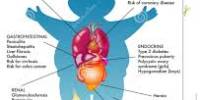
The urinary tract system is comprised of many structures that are involved in the outflow of urine from the body for excretion, and if there is a blockage at any point, it can lead to obstructive uropathy.
It can occur in both males and females and can develop at any age. However, those over 60 are considered to be at higher risk. Age is a significant risk factor owing to the fact that many processes that can possibly obstruct urine flow occur more frequently at advanced ages.
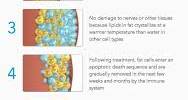
Causes, Sign, and Symptom of Obstructive Uropathy: Obstructive uropathy occurs when urine cannot drain through the urinary tract. Urine backs up into the kidney and causes it to become swollen. This condition is known as hydronephrosis.
Obstruction may occur due to the following:
- Bladder stones
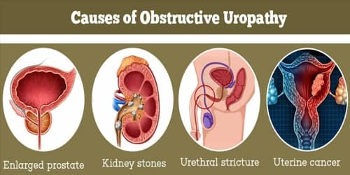
- Kidney stones
- Benign prostatic hyperplasia (enlarged prostate)
- Bladder or ureteral cancer
- Colon cancer
- Cervical cancer
- Uterine cancer
- Metastatic cancer
- Scar tissue that occurs inside or outside of the ureters
- Problems with the nerves that supply the bladder (neurogenic)
- Blood clots
- Pelvic fracture
- Disease of the digestive tract
Obstructive uropathy can affect one or both kidneys. It can occur suddenly or be a long-term problem.
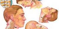
Symptoms, less likely in chronic obstruction, are pain radiating to the T11 to T12 dermatomes, anuria, nocturia, or polyuria. Fever, nausea, and vomiting are also common symptoms of obstructive uropathy. People may experience swelling or tenderness in the kidneys as urine flows backward into their organs.
Symptoms to look for include:
- difficulty passing urine
- a slowed stream, sometimes described as a “dribble”
- a frequent urge to urinate, especially at night (nocturia)
- the feeling that people’s bladder isn’t empty
- decreased urine output
- blood in people’s urine
People may have a decrease in the amount of urine they expel if just one of their kidneys is blocked. Usually, both kidneys need to be blocked to impact urine output.

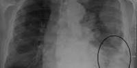
Diagnosis and Treatment of Obstructive uropathy: In almost all cases, additional diagnostic tests will have to take place to get a better idea of where the obstruction is in the urinary tract. But first, some general tests such as a urinalysis and bloodwork will be taken to look for obstructive uropathy clues.
Diagnosis is based on results of bladder catheterization, ultrasonography, CT scan, cystourethroscopy, or pyelography, depending on the level of obstruction.
A bladder catheterization is often done if urine output is diminished if there is a suprapubic pain, or if there is a distended bladder.
A bladder catheterization is often done if urine output is diminished if there is a suprapubic pain, or if there is a distended bladder.
Other diagnostic tests for the diagnosis of obstructive uropathy may include:
- Helical CT
- MRI
- IV pyelography (IVP)
- Invasive pyelography
Treatment, depending on the cause, may require prompt drainage of the bladder via catheterization, medical instrumentation, surgery (e.g., endoscopy, lithotripsy), hormonal therapy, or a combination of these modalities.
Treatment of the obstruction at the level of the ureter:
- Surgery – A surgeon will remove masses such as cancerous tumors, polyps, or scar tissue that forms in and around the patient’s ureters. Once they clear the blockage from the affected ureter, urine can flow freely into their bladder.
- Less invasive treatment – laparoscopic correction.
- Minimal invasive treatment – Overtoom procedure: dilatation with cutting balloon catheter (e.g., Boston Scientific) followed by an introduction of the pyeloplasty balloon catheter. This balloon is inflated with pure contrast agent via the pusher and remains in situ in the ureter to keep the previously treated stricture dilated while the expanded urothelium heals. Urine can drain through the central channel of this catheter.
Fetal treatment is usually performed only when the baby’s kidneys appear to be irreversibly damaged. Most often, doctors can repair kidney function and blocked ureters after the baby is born.
Information Source: