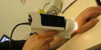
The researchers demonstrated a magnet-based system that can track muscle location and could be used to assist people in controlling prosthetic limbs and other wearable robotic devices. MIT researchers have developed a sophisticated method for monitoring muscle movements using a simple set of magnets, which they hope will make it easier for people with amputations to control their prosthetic limbs.
The researchers demonstrated the accuracy and safety of their magnet-based system, which can track the length of muscles during movement, in a new set of papers. Animal studies suggest that this strategy could be used to help people with prosthetic devices control them in a way that is more similar to natural limb movement.
“These recent results show that this tool can be used outside the lab to track muscle movement during natural activity,” says Cameron Taylor, an MIT research scientist and co-lead author of both papers.
In one of the studies, the researchers demonstrated that they could accurately measure the lengths of turkey calf muscles as the birds ran, jumped, and performed other natural movements. In the other study, they demonstrated that when small magnetic beads were implanted in muscle, they did not cause inflammation or other negative effects.
We’re able to provide the muscle-length tracking functionality of the room-sized X-ray equipment using a much smaller, portable package, and we’re able to collect the data continuously instead of being limited to the 10-second bursts that fluoromicrometry is limited to.
Hugh Herr
“I am very excited for the clinical potential of this new technology to improve the control and efficacy of bionic limbs for persons with limb-loss,” says Hugh Herr, a professor of media arts and sciences, co-director of the K. Lisa Yang Center for Bionics at MIT, and an associate member of MIT’s McGovern Institute for Brain Research.
Herr is a senior author of both papers, which appear today in the journal Frontiers in Bioengineering and Biotechnology. Thomas Roberts, a professor of ecology, evolution, and organismal biology at Brown University, is a senior author of the measurement study.
Tracking movement
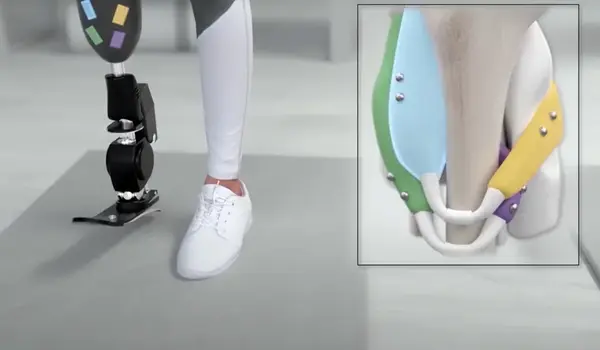
Powered prosthetic limbs are currently controlled using a technique known as surface electromyography (EMG). Electrodes attached to the skin’s surface or surgically implanted in the residual muscle of the amputated limb measure electrical signals from a person’s muscles, which are fed into the prosthesis to help it move in the manner intended by the person wearing the limb. However, that approach ignores any information about muscle length or velocity, which could help to improve the accuracy of prosthetic movements.
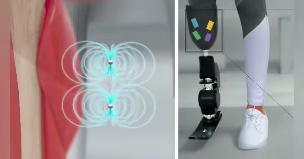
Several years ago, the MIT team began working on a novel way to perform those kinds of muscle measurements, using an approach that they call magnetomicrometry. This strategy takes advantage of the permanent magnetic fields surrounding small beads implanted in a muscle. Using a credit-card-sized, compass-like sensor attached to the outside of the body, their system can track the distances between the two magnets. When a muscle contracts, the magnets move closer together, and when it flexes, they move further apart.
When the beads were implanted in the calf muscles of turkeys, the researchers demonstrated that this system could be used to accurately measure small ankle movements in a study published last year. The researchers set out to see if the system could make accurate measurements during more natural movements in a nonlaboratory setting in one of the new studies.
To accomplish this, they built an obstacle course consisting of ramps for the turkeys to climb and boxes for them to jump on and off of. The researchers tracked muscle movements with their magnetic sensor during these activities and discovered that the system could calculate muscle lengths in less than a millisecond.
They also compared their data to measurements taken using a more traditional approach known as fluoromicrometry, a type of X-ray technology that requires much larger equipment than magnetomicrometry. The magnetomicrometry measurements varied from those generated by fluoromicrometry by less than a millimeter, on average.
“We’re able to provide the muscle-length tracking functionality of the room-sized X-ray equipment using a much smaller, portable package, and we’re able to collect the data continuously instead of being limited to the 10-second bursts that fluoromicrometry is limited to,” Taylor says.
Biocompatibility
The researchers focused on the biocompatibility of the implants in the second paper. They discovered that the magnets did not cause tissue scarring, inflammation, or other negative effects. They also demonstrated that the implanted magnets did not alter the turkeys’ gaits, implying that they did not cause discomfort. The biocompatibility study was co-authored by William Clark, a postdoc at Brown.
The researchers also demonstrated that the implants remained stable for the duration of the study, eight months, and did not migrate toward each other as long as they were implanted at least three centimeters apart. The beads, which are made of a magnetic core coated in gold and a polymer called Parylene, could be implanted and remain in tissue indefinitely, according to the researchers.
“Magnets do not require an external power source, and after being implanted into the muscle, they can maintain the full strength of their magnetic field for the duration of the patient’s life,” Taylor explains.
The researchers will now seek FDA approval to test the system on people who have prosthetic limbs. They hope to use the sensor to control prostheses in the same way that surface EMG is currently used: measurements of muscle length will be fed into the control system of a prosthesis to help guide it to the position that the wearer intends.
Magnet-based #sensors for tracking muscle length could help people with prosthetic #devices.https://t.co/GNWxLb4AkP
— ACS Sensors (@ACS_Sensors) October 31, 2022