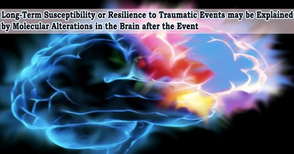
Social withdrawal is a classic sign of PTSD, and researchers trying to understand why have found laboratory evidence that while stress hormone levels rise steadily right after a traumatic incident, there may be completely different effects on different sections of the brain later on.
Some rodent models exhibit the expected short-term increase in the excitability of neurons in regions of their brain important for memory and how they perceive their environment, as part of the natural instinct to fight or flee, in response to a significant stressor and the ensuing surge of stress hormones.
Other genetically identical mice instead experience a decrease in neuron excitability in this key area called the dorsal hippocampus, Dr. Chung Sub Kim, neuroscientist at the Medical College of Georgia at Augusta University, and his colleagues report in the journal Molecular Psychiatry.
Human PTSD has been associated to low levels of neuronal activity in the hippocampus, and comprehensive brain imaging of PTSD patients shows structural and functional alterations in important brain regions, including the hippocampus.
In the hippocampus, which is abundantly expressed, cortisol-releasing glucocorticoid receptors have been found to be more highly expressed in PTSD patients than in controls during stressful events.
“We are trying to answer the question as to why hippocampal activity is decreased in PTSD or depressed patients,” Kim says.”We know it happens, but the mechanism we don’t know.”
They have discovered, among other things, that certain mice, like some people, just seem more susceptible to the long-term effects of a significant and/or chronic stressor, and that both their behavior and internal biochemical reaction to stress are distinct from their more resilient colleagues.
There are clearly physical differences in the response to stress in the two mice that correlate with their behavior.
Dr. Chung Sub Kim
“One was affected directly by stress, and another not so much,” Kim says.
The researchers set up a scenario where a male mouse, which is naturally aggressive, established his area, then repeatedly assaulted another mouse who dared to enter that region in order to simulate stressful situations like being bullied or being the victim of an armed robbery.
Again, somewhat like human victims, some of the mice did not seem phased after the attack, rather were still naturally inquisitive about the other mouse; while the susceptible mouse clearly avoided The aggressor.
Kim and his colleagues discovered elevated expression of stress hormone receptors on neurons in the CA1 area of the dorsal hippocampus in the “susceptible” animals. A natural regulator of neuron activity and connectivity that is already present in large levels in the hippocampus, the protein HCN1, appears in turn and maybe counterintuitively to be made possible by those many receptors.
HCN1 is a major research focus for Kim, who has evidence that even a single episode of significant stress can further increase HCN1 expression in the CA1 region of the dorsal hippocampus and bring neuron excitability down. Also increased in the susceptible rodents wasthe protein TRIP8b, which regulates HCN channel levels. “Stress changes everything,” Kim says.
The researchers discovered that this cascade also led to a rise in another naturally occurring mechanism for tamping down activity, known as hyperpolarization-activated current. This process was known to be heightened by stress, but how exactly was unknown. Again, the changes were specific to the dorsal in humans the back part portion of the hippocampus.
These levels that reduced neuron excitability were still present months later, and the sensitive mice continued to avoid the hostile male mouse. Even after the direct injection of a stress hormone to the neurons, which should once more raise neuron excitability, the reduced neuron excitability persisted.
In addition to having poor spatial working memory, the susceptible mice also had difficulty remembering where they had put their vehicle keys and how to get to work.
The clearly different expression of the HCN1 protein in this region of the hippocampus may be the molecular mechanism driving susceptibility to social avoidance, Kim and his colleagues write.
“They have some malfunction in hippocampal information processing,” he says. Whether those changes are permanent is not certain but at three months, a long time in mouse years, they were still present: The average mouse lives maybe two to three years, while the average human in the U.S. lives into their late 70s.
But in the “resilient” mice, expression of the stress hormone receptor and HCN channel did not increase, but neuron excitability did, in the immediate aftermath of stress.
“There are clearly physical differences in the response to stress in the two mice that correlate with their behavior,” Kim says, even though you would not suspect the differences in these genetically identical rodents.
The researchers add that further research is still needed to pinpoint why some mice are resilient to emotional trauma while others are more vulnerable to it.
According to Kim and his colleagues, the ventral hippocampus is more associated with emotional reactions like fear whereas the dorsal hippocampus is more associated with learning and memory in mice. Comparatively speaking, the dorsal hippocampus has less neuron excitability and is clearly the most reactive to chronic stress.
Many physiological functions, including sleep and wake states, taste and fear learning, include HCN channels. Kim’s research and those of others has revealed evidence of a connection between HCN channels and mental illnesses like depression and anxiety.
Cortisol and adrenaline are also released by the adrenal gland in reaction to a dangerous circumstance. By making modifications like raising glucose levels, which your body utilizes as fuel, and suppressing operations like digestion and reproduction, which are not deemed necessary at that time, the increase helps the body get ready for the so-called protective flight or fight response.
The amygdala, which aids in remembering and storing emotions like anger, fear, and sadness as well as recognizing threat, and the medial prefrontal cortex, which is thought to be important to cognitive functions like attention, habit formation, and long-term memory, have both been shown to change in individuals with PTSD.