Chest Radiograph
Definition
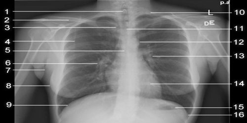
Chest radiograph, which is also called a chest X-ray (CXR), or chest film is a picture of the chest that shows our heart, lungs, airway, blood vessels, and lymph nodes. A chest X-ray also shows the bones of our spine and chest, including our breastbone, our ribs, our collarbone, and the upper part of our spine. A chest X-ray is the most common imaging test or X-ray used to find problems inside the chest. Chest radiographs are the most common film taken in medicine.
Doctor could order a chest X-ray for a variety of reasons, including to assess injuries resulting from an accident or to monitor the progression of a disease, such as cystic fibrosis. People might also need a chest X-ray if they go to the emergency room with chest pain or if they have been involved in an accident that included force to their chest area.
A chest X-ray (or Chest radiographs) is an easy, quick, and effective test that has been useful for decades to help doctors view some of our most vital organs. The mean radiation dose to an adult from a chest radiograph is around 0.02 mSv (2 mrem) for a front view (PA or posterior-anterior) and 0.08 mSv (8 mrem) for a side view (LL or latero-lateral). Together, this corresponds to a background radiation equivalent time of about 10 days.
Performed and Views of Chest Radiographs
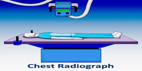
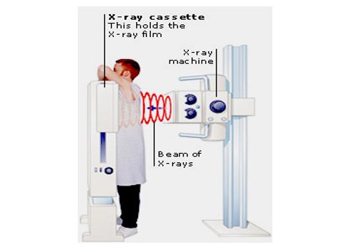
The X-ray (or Radiographs) occurs in a special room with a movable X-ray camera attached to a large metal arm. Patient will stand next to a “plate.” This plate may contain X-ray film or a special sensor that records the images on a computer. They will wear a lead apron to cover their genitals. This is because their sperm (men) and eggs (women) could be damaged from the radiation. While the images are taken, patient will need to hold their breath so that their chest stays completely still. If they move, the images might turn out blurry. As the radiation passes through their body and onto the plate, denser materials, such as bone and the muscles of their heart, will appear white.
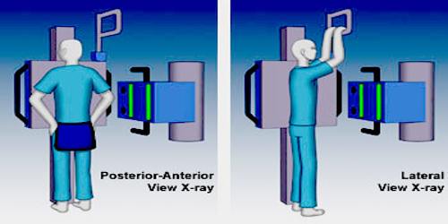
Different views, which is also known as projections of the chest can be obtained by changing the relative orientation of the body and the direction of the x-ray beam. The most common views are posteroanterior, anteroposterior, and lateral.
- Posteroanterior (PA) views: In a posteroanterior (PA) view, the x-ray source is positioned so that the x-ray beam enters through the posterior (back) aspect of the chest, and exits out of the anterior (front) aspect where the beam is detected. To obtain this view, the patient stands facing a flat surface behind which is an x-ray detector. A radiation source is positioned behind the patient at a standard distance (most often 6 feet, 1,8m), and the x-ray beam is fired toward the patient.
- Anteroposterior (AP) views: In anteroposterior (AP) views, the positions of the x-ray source and detector are reversed: the x-ray beam enters through the anterior aspect and exits through the posterior aspect of the chest. In this situation, mobile X-ray equipment is used to obtain a lying down chest x-ray (also known as a “supine film”). As a result, most supine films are also AP.
- Lateral views: In lateral views of the chest are obtained in a similar fashion as the posteroanterior views, except in the lateral view, the patient stands with both arms raised and the left side of the chest pressed against a flat surface.
Required projections can vary by country and hospital, typically an erect postero-anterior (PA) projection is first preference, and if this is not possible then an antero-posterior view will be taken. Lateral decubitus may be used for visualization of air-fluid levels if an upright image cannot be obtained. Anteroposterior (AP) Axial Lordotic projects the clavicles above the lung fields, allowing better visualization of the apices, which is extremely useful when looking for evidence of primary tuberculosis.
Some Common Uses of Chest Radiographs
The chest x-ray (or Chest Radiographs) is performed to evaluate the lungs, heart and chest wall.
A chest x-ray is typically the first imaging test used to help diagnose symptoms such as:
- breathing difficulties.
- a bad or persistent cough.
- chest pain or injury.
- fever.
Physicians use the examination to help diagnose or monitor treatment for conditions such as:
- pneumonia.
- heart failure and other heart problems.
- emphysema
- lung cancer.
- positioning of medical devices.
- fluid or air collection around the lungs.
- other medical conditions.
Risk: There is always a slight chance of damage to cells or tissue from radiation, including the low levels of radiation used for this test. But the chance of damage from the X-rays is usually very low compared with the benefits of the test.
Reference: