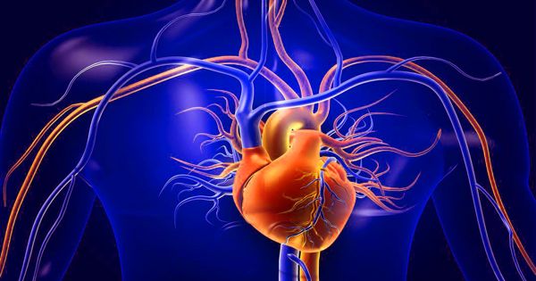
Cardiac muscle, also known as the myocardium, is a type of muscle tissue found exclusively in the heart. It is one of three types of vertebrate muscle tissues, along with skeletal and smooth muscle. It is an involuntary, striated muscle that makes up the majority of the heart’s wall. It is in charge of the contraction and relaxation of the heart chambers, which allows blood to be pumped throughout the circulatory system.
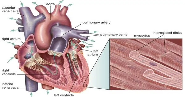
The coronary circulation supplies blood to the cardiac muscle (myocardium), which forms a thick middle layer between the outer layer of the heart wall (pericardium) and the inner layer (endocardium). Cardiac muscle differs in structure and function from other types of muscle tissue, such as skeletal and smooth muscle. It is made up of individual cardiac muscle cells connected by intercalated discs and surrounded by collagen fibers and other extracellular matrix substances.
Cardiac muscle contracts similarly to skeletal muscle, but with some significant differences. The release of calcium from the cell’s internal calcium store, the sarcoplasmic reticulum, is triggered by electrical stimulation in the form of a cardiac action potential. Excitation-contraction coupling occurs when calcium levels rise and myofilaments slide past each other in the cell. Cardiomyopathies, or diseases of the heart muscle, are extremely serious. Angina and myocardial infarction are examples of ischemic conditions caused by a restricted blood supply to the muscle.
Here are some key features of cardiac muscle:
- Striated Appearance: Cardiac muscle, like skeletal muscle, is striated, which means it has a striped appearance when viewed under a microscope. The organized arrangement of contractile proteins within muscle cells causes this striation.
- Involuntary Control: Cardiac muscle is controlled involuntarily, which means it contracts without conscious effort. The autonomic nervous system, specifically the cardiac conduction system, regulates contraction.
- Intercalated Discs: Intercalated discs, which are specialized structures that allow cells to communicate quickly, connect these cells. Gap junctions in intercalated discs allow for coordinated contraction of the entire heart muscle.
- Single Nucleus: In contrast to skeletal muscle, each cardiac muscle cell typically has a single nucleus.
- Highly Mitotic: Although cardiac muscle cells do not divide as actively as cells in some other tissues, they have some capacity for regeneration, primarily through the division of existing cells.
- Endurance: It is designed for endurance and continuous rhythmic contractions. The heart beats consistently throughout a person’s life, and cardiac muscle is adapted to withstand fatigue.
- Aerobic Metabolism: It generates the energy required for contraction primarily through aerobic metabolism (oxygen-dependent). In contrast, skeletal muscle can also use anaerobic metabolism for short bursts of energy.
Coordination of cardiac muscle contraction is critical for maintaining blood circulation and supplying oxygen and nutrients to the body’s tissues. The heart is divided into four chambers: two atria (upper chambers) and two ventricles (lower chambers), all of which are made up of cardiac muscle tissue. The heart’s contraction is controlled by a complex interplay of electrical signals and hormonal influences. Heart muscle dysfunction can result in a variety of cardiovascular conditions, including heart failure and arrhythmias.