Rare eye disorders can be challenging to study due to the small number of affected individuals and the limited amount of available data. However, large-scale studies can enable new insights into these conditions by pooling data from multiple sources. One example of a large-scale study on rare eye disorders is the UK Biobank Eye and Vision Consortium. This consortium is a collaboration between researchers from 12 UK institutions and the UK Biobank, a large-scale biomedical database with data from over 500,000 participants.
Researchers examined image and genomic data from the UK Biobank to gain insight into rare eye diseases. These include retinal dystrophies, which are a group of inherited retinal disorders that are also the leading cause of blindness certification in working-age adults.
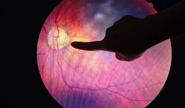
The retina is located behind the pupil of the eye. It is a layered tissue that receives light and converts it into a signal that the brain can understand. Each retinal layer contains various cell types that each play a distinct role in the light conversion process.
The researchers focused on photoreceptor cells (PRCs), which are light-detecting cells found in the retina, for this study, which was published in the journal PLOS Genetics. These cells can be imaged non-invasively using optical coherence tomography (OCT), a service now widely available in many opticians. Researchers were able to generate the largest genome-wide association study of PRCs using OCT image data and genomic data stored in the UK Biobank.
We had access to coupled images and genotype data on a scale never seen before in a study of this kind. We were able to identify genetic links to rare retinal dystrophies thanks to access to this massive amount of data. This work has opened up new research avenues and raised new questions about rare retinal dystrophies.
Hannah Currant
Rare retinal dystrophies
Rare diseases of the retina are frequently caused by inherited mutations in genes expressed by PRCs. These mutations cause the retina to function incorrectly, resulting in sight impairment or even blindness. Although these individual diseases are rare, together they are the leading cause of blindness in working-age adults.
“We had access to coupled images and genotype data on a scale never seen before in a study of this kind,” said Hannah Currant, a former Ph.D. student at EMBL’s European Bioinformatics Institute (EMBL-EBI) and Postdoctoral Fellow at the Novo Nordisk Foundation Center for Protein Research (CPR) at the University of Copenhagen. “We were able to identify genetic links to rare retinal dystrophies thanks to access to this massive amount of data. This work has opened up new research avenues and raised new questions about rare retinal dystrophies.”
Linking genotype and phenotype
OCT generates high-resolution images that can be used to identify the retina’s various layers and structures. In the clinic, these images are frequently used to aid in the diagnosis of eye disorders. The researchers used OCT images and the corresponding genomic and medical information from over 30,000 participants stored in the UK Biobank for this study.
“The UK Biobank is a rich, invaluable resource with enormous potential for genomic medicine,” Ewan Birney, Deputy Director General of the European Molecular Biology Laboratory, said (EMBL). “There is so much potential waiting to be released from the data stored there, which allows us to understand human biology as well as how and when things go wrong in disease.”
Driving genomic medicine
The researchers used genome-wide association studies (GWAS) on UK Biobank data to look for genetic variations associated with differences in PRC layer thickness. This led them to discover genomic variations linked to the thickness of one or more of the PRC layers, including those previously linked to known eye diseases. The newly discovered genomic associations are saved and can be accessed freely via the GWAS Catalog.
Some of these genetic variants were already known to be associated with eye diseases, but surprisingly, a number of relatively common genetic variants were found near genes known to cause rare genetic eye diseases when disrupted. In one case, the researchers were able to investigate how combinations of common variants near genes implicated in rare eye diseases alter the structure of the retina. This increases confidence when investigating specific rare disease collections to see how these common variants may impact disease.
“Systematic bioinformatic analysis of large-scale participant data cohorts is driving the future of genomic medicine,” said Omar Mahroo, Professor of Retinal Neuroscience at University College London and Consultant Ophthalmologist at Moorfields Eye Hospital. “Having access to these data and being able to make these connections between disease phenotypes and genetic variation will open many new opportunities for modern disease diagnosis and therapeutics.”
EMBL’s priority is to develop a molecular understanding of how the human ecosystem – the social, physical, and biological factors we are exposed to throughout our lives – interacts with our genetic makeup to influence our health. Learn more about how EMBL researchers will use the unprecedented depth and breadth of emerging human cohort data to improve our understanding of human diseases.