The smart application of a frequent occurrence in quantum physics has greatly improved the resolution of light microscopes.
It is feasible to stare at fragile things more carefully than ever before by sending entangled light along separate channels and recombining their waves, thus doubling their resolution without the normal complexity of drastically raising the light’s intensity.
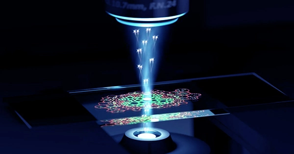
It’s known as quantum microscopy by coincidence (QMC), and it was created by researchers at the California Institute of Technology (Caltech) in the United States. They believe it’s especially well suited to examining tissues and biomolecules to detect illness or analyze its spread.
“The combination of improved speed, enhanced contrast-to-noise ratio, more robust stray light resistance, super resolution, and low-intensity illumination empowers QMC toward bioimaging,” the researchers write in their new work.
Prior to being noticed, quantum entanglement defines correlations that exist between things that have a common past. Particles can be mathematically linked in a multitude of ways, much as two shoes purchased in a store are correlated to suit a right foot and a left foot.
Shoes and electrons, for example, do not completely settle into any of those states until they are detected in a quantum system. They are just possibilities that may be expressed as a wave of maybes.
The particles involved in QMC were photons, or light particles, which become biphotons after being entangled in a pair.
This was accomplished using a unique crystal composed of -barium borate (BBO). When laser light passes through the crystal, only a very tiny proportion of the photons – around one in a million – are transformed into biphotons. The biphotons were then separated once more by the researchers using a network of mirrors, lenses, and prisms.
One photon is delivered through the substance being investigated, while the other is examined. Because they are entangled, the correlations observed in any photon will provide information about its partner’s path. It serves as the foundation for another relatively new technique known as ghost imaging.
This interwoven dual-action, however, has another surprise up its sleeve. Biphotons have twice the momentum of photons, resulting in halved wavelengths. A light microscope with half the wavelength of light has a better resolution. Light with shorter wavelengths typically carries more energy, which can eventually harm the cells being researched.
Consider the contrast between long-lasting radio waves and the more potent, short-lasting ultraviolet (UV) radiation that may destroy DNA and cause sunburn.
While the entanglement mechanism essentially halves the wavelength in this example, it does not enhance the energy of the individual photons.
“Cells don’t like UV light,” explains Lihong Wang, a medical engineer at the California Institute of Technology (Caltech). “However, if we can image the cell with 400-nanometer light and achieve the effect of 200-nm light, which is UV, the cells will be happy, and we’ll get UV resolution.”
There is also space for development in this technology, such as faster imaging and the ability to entangle more photons together, boosting resolution even more. However, adding additional photons reduces the possibility of entanglement, which is already one in a million, even further.
Because interactions with the environment may readily shatter entanglement, increasing the number of photons in a system increases the possibility that individual photons will interact with the environment rather than with one another.
While biphoton imaging has been attempted in the past, the researchers behind the new setup have made significant changes and tested it realistically, making it one of the most promising approaches of its sort.
“We developed what we believe is a rigorous theory, as well as a faster and more accurate entanglement-measurement method,” Wang explains. “We reached microscopic resolution and imaged cells.”