Scientists have long puzzled over how cells interact with one another, but Rutgers researchers have cracked the code using a tiny roundworm.
The research, published in Current Biology, could aid in the development of medicines for Alzheimer’s and other neurodegenerative illnesses.
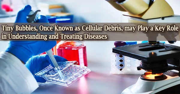
Cells communicate good and negative news to one another in a variety of ways, one of which is through tiny bubbles known as extracellular vesicles (EVs). EVs deliver useful or hazardous cargo that promotes good health or sickness and were once thought to be cellular trash.
EVs, for example, convey disease-causing proteins in the human brain that may influence the progression of Alzheimer’s disease.
“Although EVs are of profound medical importance, the field lacks a basic understanding of how EVs form, what cargo is packaged in different types of EVs originating from the same or different cell types, and how different cargos influence the range of EV targeting and bioactivities,” said lead author Inna Nikonorova, a postdoctoral researcher.
Because healthy and ill cells package distinct EV cargo, EVs, which are prevalent in human fluids such as urine and blood, could be employed as biomarkers for disease in liquid biopsies.
The Rutgers team opted to investigate the unknown function of EVs in our bodies using a basic experimental animal called C. elegans, or roundworms, as well as cutting-edge genetic, molecular, biochemical, and computational technologies.
We developed an innovative method to label, track and profile EVs using genetically encoded, fluorescent-tagged EV cargo and conducted large-scale isolation and protein profiling. Using this strategy, we discovered four novel cilia EV cargo. Combined, these data indicate that C. elegans produces a complex and heterogeneous mixture of EVs from multiple tissues in living animals and suggests that these environmental EVs play diverse roles in animal physiology.
Inna Nikonorova
Large-scale identification research led by Maureen Barr, a professor in the Department of Genetics, and Nikonorova discovered 2,888 EV cargo candidates.
Nikonorova concentrated on EVs produced by cilia, the cellular antennae that transmit and receive signals for intercellular communication, due to the relevance of EVs in the human nervous system.
The researchers focused on EV cargo produced by nerve cells and discovered that EVs transport RNA-binding proteins as well as RNA, which can be observed in the COVID-19 mRNA vaccine’s function in therapeutic therapies.
Nikonorova and Barr theorized that neurons wrap RNA-binding proteins and RNA into EVs to facilitate cell-to-cell and animal-to-animal communication. For generating tailor-made EVs for RNA-based therapeutics, a basic understanding of EV-RNA biology is critical.
“We developed an innovative method to label, track and profile EVs using genetically encoded, fluorescent-tagged EV cargo and conducted a large-scale isolation and protein profiling,” Nikonorova said.
“Using this strategy, we discovered four novel cilia EV cargo. Combined, these data indicate that C. elegans produces a complex and heterogeneous mixture of EVs from multiple tissues in living animals and suggests that these environmental EVs play diverse roles in animal physiology.”
Understanding EV-mediated RNA communication will be the focus of future research in the Barr lab. The National Institute of Neurological Disorders and Stroke, as well as the National Institute of Diabetes and Digestive and Kidney Diseases, support Barr’s research.