According to researchers, an enzyme that is being tested as a potential treatment for some cancers may also be able to lessen the severe eyesight loss that diabetes and preterm delivery can cause.
Inflammation is seen as a cancer hallmark. It is widespread in both of these potentially blinding eye disorders because it causes the formation of new blood vessels to improve oxygen delivery, but these new blood vessels frequently clog the vision pathway and become leaky, which leads to swelling and further impairs vision.
There is growing evidence, according to researchers at the Medical College of Georgia, that increasing the availability of the enzyme arginase 1, or A1, can help reduce these unhealthy reactions and stop a physiological process that normally promotes persistent, high levels of destructive inflammation in both diabetic retinopathy and retinopathy of prematurity. These studies were recently published in the journals Cell Death and Disease and Cells.
Making less of the amino acid L-arginine accessible is essential to the process. For instance, in diabetes, elevated levels of blood sugar and lipids, in addition to oxidative stress, increase the expression of inducible nitric oxide synthase, or iNOS, which uses the amino acid L-arginine to aid in the production of additional inflammation and to advance the course of the disease.
In order for it to function, iNOS is intended to increase in response to an infection, and then high-expressing A1 cells should enter the scene to switch iNOS down and reduce inflammation.
“That’s because A1 competes with iNOS for L-arginine. They theorized that more A1, which actually breaks down L-arginine into two products, would make less L-arginine available to “feed this (unhealthy) iNOS explosion,” and help tamp down the vicious cycle of inflammation and related damage,” says Dr. William Caldwell, pharmacologist and chair emeritus of the MCG Department of Pharmacology and Toxicology.
AI’s anti-inflammatory power is exhibited in the way it can keep those immune cells, called macrophages, which can both promote and reduce inflammation, from becoming too proinflammatory.
Dr. William Caldwell
“If you reduce L-arginine levels, iNOS cannot work. This will make things better,” Caldwell says.
Nitric oxide, a potent blood vessel dilater produced by iNOS, is healthy for blood vessels and blood pressure when it is produced at low levels by the endothelial cells that line blood arteries. This is a perfect illustration of how everything in the body is about balance. This NO has the added benefit of being anti-inflammatory.
“But the “toxic” high levels produced by iNOS result in production of more reactive oxygen species, which feeds inflammation, which basically “rusts” whatever tissue it touches,” Caldwell says.
They discovered that giving A1 three times a week for two weeks to an obese, diabetic mouse that constantly feeds and develops a condition resembling early-stage diabetic retinopathy enhances the mice’ visual acuity and helps them discern between different colors of darkness.
“The mice had less oxidative stress and inflammation in the retina, restoration of the protective blood retinal barrier to help avoid leakage of tiny capillaries and the swelling and damage that would follow, and reduced progression of diabetic retinopathy,” says Dr. Ruth B. Caldwell, cell biologist in the MCG Vascular Biology Center.
In contrast, they have demonstrated that mice that express half as much A1 as normal do so with greater aberrant blood vessel growth and retinal damage, they write.
According to William Caldwell, when A1 is totally eradicated, mice and humans would succumb to ammonia poisoning, as it constantly works in the liver to assist eliminate ammonia, which is created when proteins, a fundamental component of the body, are broken down.
That is how L-arginine, which A1 cleaves or cuts to produce urea, an ammonia form that may be removed in the urine, and l-ornithine, which is essential for typical physiological processes like cell proliferation and collagen synthesis, is occupied so much by A1.
Additionally, the MCG researchers have demonstrated that A1 is normally present in immune cells and retinal cells of mice with oxygen-induced retinopathy, a popular model for the apoptotic blood vessel development that takes place in retinopathy of prematurity.
Because diabetic mice don’t live long enough to acquire full-blown eye illness, the model is also used to imitate some elements of diabetic retinopathy. Additionally, they discovered A1 in blood samples from young children with retinopathy of prematurity as well as in the retinas of people with diabetic retinopathy.
“AI’s anti-inflammatory power is exhibited in the way it can keep those immune cells, called macrophages, which can both promote and reduce inflammation, from becoming too proinflammatory,” Ruth Caldwell says.
She points out that numerous anti-inflammatory drugs and antioxidants have been tried and failed to treat patients with these conditions. “What we think is happening is this pegylated A1 is reducing within these immune cells the ability to make inducible nitric oxide synthase in big amounts. We think that is the deal.”
Similar to research on cancer, which cannot grow and survive without L-arginine, A1 generally degrades within minutes, therefore they utilized a synthetic pegylated, or stable, form that lasts for days instead.
According to the Caldwells, new, easily accessible treatments are required to help prevent the vision consequences of both type 1 and type 2 diabetes as well as retinopathy of prematurity.
According to their research, A1 can permeate the eye in conditions when the blood-retinal barrier is compromised. This eliminates the need for eye injections and should make treatment much more widely available.
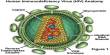
Anti-VEGF (vascular endothelial growth factor) therapy, for example, used to also combat the growth of abnormal and leaky blood vessels, requires injections into the back of the eye.
The Caldwells are co-corresponding authors on the two new papers. Dr. Abdelrahman Y. Fouda, Ruth Caldwell’s former postdoctoral fellow who is now on the faculty of the University of Arkansas for Medical Sciences College of Medicine, is first author on the Cell Death and Disease paper. Dr. Ammar A. Abdelrahman, a postdoc in William Caldwell’s lab, is first author on the Cells paper.
The scientific team has already demonstrated that administering pegylated A1 improves protection in ischemia/reperfusion injury where oxygen supply is temporarily lost, as with a knife wound, then the blood supply is restored in major optic nerve trauma where a clot typically cuts off the blood and oxygen supply to a part of the brain and after an ischemic stroke, which is by far the most common type of stroke.
The research projects were supported by the National Eye Institute, the James and Jean Culver Vision Discovery Institute at Augusta University and the Department of Veterans Affairs.