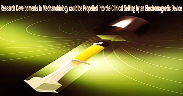
An electromagnetic device, such as a magnetic tweezers or a magnetic twisting cytometry, can be used to apply precise and controlled mechanical forces to living cells in order to study the effects of mechanical stress on cell behavior and physiology.
Researchers claim that a novel electromagnetic tool has set a new bar for precision in the field of mechanobiology by enabling high-precision measurements of a variety of soft biological tissues. The technique makes it possible to mechanically examine tissues the size of human biopsy samples, which makes it very pertinent for research on human disease.
The soft tissues in the body have a wide range of mechanical characteristics, like stiffness and strength, which are essential for them to function. For instance, tendons are substantially more rigid to transmit force from muscle to bone so that we can move, whereas the tissues of the gastrointestinal system are soft to allow for the transit and digestion of food.
Biology and medicine stand to gain significantly from the capacity to precisely evaluate the mechanical characteristics of these tissues, which are prone to alter throughout developmental processes or as a result of disease. The precision and reliability of the methods used to measure these qualities are still limited at this time.
Our study demonstrated the enhanced reliability of the electromagnetic device, yielding errors in the stress-strain response below 15% a level of accuracy not seen before. We hope that this device may eventually become the new standard in the tissue biomechanics field, providing a standardised dataset for the characterisation of mouse and human soft tissue mechanics across the board.
Dr. Adrien Hallou
New research involving researchers from the University of Cambridge and the MIT Institute for Medical Engineering and Science (IMES) has resulted in a device that relies on magnetic actuation and optical sensing, thus potentially allowing for live imaging of the tissue under an inverted microscope. The behavior of the tissue under mechanical pressures at the cellular and molecular levels can be better understood in this way. The results are reported in the journal Science Advances.
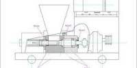
The tissue specimen is positioned on the apparatus, and an electromagnet pulls on it while an optical system measures any changes in the specimen’s size or form.
“One of the most critical requirements for mechanical testing of soft biological tissues is the need to mimic the biological specimen’s physiological conditions (e.g., temperature, nutrients) as closely as possible, in order to keep the tissue alive and preserve its biomechanical properties,” said Dr. Thierry Savin, Associate Professor in Bioengineering, who led the research team.
“To this end, we designed a transparent mounting chamber to measure the mechanical properties of tissues at the millimetre scale in their native physiologic and chemical environment. The result is a more versatile, precise and robust device that shows high reliability and reproducibility.”
The researchers studied the biomechanics of a mouse esophagus and its constituent layers to directly evaluate the effectiveness of their electromagnetic device. The muscular tube that connects the throat to the stomach is called the esophagus, and it has several tissue layers.
The tool was employed by the researchers to carry out the initial biomechanical analysis of each of the three distinct layers of mouse esophagus tissue. According to their research, the esophagus functions like a three-layer composite material, much like those that are frequently utilized in a variety of technical applications. These are, as far as the researchers are aware, the first findings regarding the mechanical characteristics of each particular layer of the esophagus.
“Our study demonstrated the enhanced reliability of the electromagnetic device, yielding errors in the stress-strain response below 15% a level of accuracy not seen before,” said Dr. Adrien Hallou, Postdoctoral Fellow at the Wellcome Trust/Cancer Research UK Gurdon Institute.
“We hope that this device may eventually become the new standard in the tissue biomechanics field, providing a standardised dataset for the characterisation of mouse and human soft tissue mechanics across the board.”
Luca Rosalia, PhD candidate at IMES, added: “Through analysis of the biomechanics of healthy tissues and their changes as they occur during disease, our device could eventually be used to identify alterations in tissue properties that are of diagnostic relevance, therefore becoming a valuable tool to inform clinical decisions.”